The topographical anatomy of the head and neck of the horse / [O. Charnock Bradley].
- Orlando Charnock Bradley
- Date:
- 1923
Licence: In copyright
Credit: The topographical anatomy of the head and neck of the horse / [O. Charnock Bradley]. Source: Wellcome Collection.
38/248 page 22
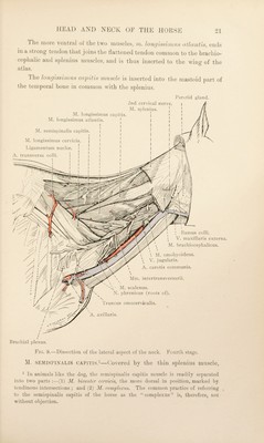
![the semispinahs capitis will be easily recognised by its great size and strength and by the fact that it is crossed obliquely by four or five tendinous intersections. The origin of the muscle is from the spinous processes of the second, third and fourth thoracic vertebra^ (in common with the splenius), the transverse processes of the first six or seven thoracic and the articular processes of the last five or six cervical vertebrae. A strong tendon begins over the atlas and is inserted into the occipital bone close to the attachment of the ligamentum nuchae. Dissection.—Make a transverse incision through the semispinahs capitis about the middle of the neck, and sever its origin from the thoracic spinous processes. This will allow' the twm parts of the muscle to be turned downwards, and wall expose certain underlying muscles. M. SPINALIS (et semispinalis) cervicis.—The origin (from the first four thoracic spinous processes) and the greater part of the extent of the spinalis dorsi et cervicis belongs to the dissector of the thorax, but its insertion into the spinous processes of the last four or five cervical vertebrae should be examined during the present dissection. This insertion lies medial to the semispinalis capitis. The dorsal branches of the cervical nerves from the third to the eighth should be examined before any further dissection is conducted. The branches of the third, fourth, fifth and sixth nerves form the dorsal cervical 'plexus, which lies between the semispinalis capitis and the ligamentum nuchae. From this plexus cutaneous nerves pass to the skin in the neighbourhood of the mid-dorsal line. Other filaments supply the adjacent muscles. The relatively small dorsal branches of the seventh and eighth cervical nerves pass in a dorsal direction between the multifidus cervicis and the longissimus cervicis, and end in the cervical rhomboid muscle, and the superjacent skin. M. MULTIFIDUS CERVICIS.—The multifidus muscle of the neck consists of five or six strong bundles arising in succession from the artictilar processes of the last four or five cervical and the first thoracic vertebrae. Each bundle is divisible into two sets of fibres, the more superficial of which pass obliquely in a cranial and medial direction to be inserted into the spinous process of a cervical vertebra. The deeper fibres are shorter in length and straighter in direction and are inserted into the articular process of a vertebra. The last bundle is inserted into the spinous process of the epistropheus. 1 Multifidus (from multus, mSiiiy + find ere, to cleave or split), [L.], cleft or divided into many parts.](https://iiif.wellcomecollection.org/image/b29820066_0038.jp2/full/800%2C/0/default.jpg)