The topographical anatomy of the head and neck of the horse / [O. Charnock Bradley].
- Orlando Charnock Bradley
- Date:
- 1923
Licence: In copyright
Credit: The topographical anatomy of the head and neck of the horse / [O. Charnock Bradley]. Source: Wellcome Collection.
49/248 page 33
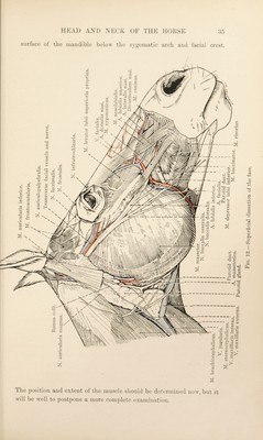
![consists of a well-marked band of fibres runnino’ obliquely, immedi- ately underneath the skin, to the angle of the mouth, where they blend with those of the buccinator and orbicular muscles. yi ORBICULARIS ocuLL —The Orbicular muscle of the eyelids consists of fibres circularly disposed so as to act as a sphincter of the palpebral Hssure. The peripheral limit of the muscle is not well defined. A few fibres are attached to the oral lachrymal tubercle of the lachrymal bone, but otherwise there is no direct attachment to bone. M. CORRUGATOR SUPERCILII.I — A feebly developed and thin muscle springs from the surface of the frontal bone and merges into the orbicular muscles in the upper eyelid. At this stage the dissector sliould make a preliminary examination of the bps and nostrils before proceeding to the removal of the skin from these structures. The two lips (labia oris) are not alike. The upper (labium superius), attached to the incisive bones, is the better developed and the more moveable. It is, moreover, marked in the middle line by a shallow and ill-defined groove, the philtrum. The lower lip (labium inferius) is connected with that part of the mandible that carries the incisor and canine teeth. Interiorly it merges into the chin (mentum), a rounded prominence composed of muscle, fat and fibrous tissue. The opening between the lips (rima oris) terminates on each side at the angle of the mouth (angulus oris) where the upper and lower lips are connected by the commissures (commissurse labiarum). The nostril (apertura nasi externa) is a crescentic opening with medial and laterial icings (alse nasi). The medial wing projects and has a basis formed by the lamina of the alar cartilage—a structure that will be exposed at a later stage of the dissection. The thinner, concave lateral wing is more flexible and consists of skin enclosing muscle and fibrous tissue. The two wings meet at a sharp upper commissure. The loiver commissure, on the contrary, is rounded, its form being governed by the curved cornu of the alar cartilage. If the finger or an instru¬ ment be thrust into the upper angle of the nostril, it does not enter the nasal cavity, but finds its way into a blind diverticulum of the nostril (di\ erticulum nasi). Ihe diverticulum, some five or six centimetres in length, extends backwards to the angle of union of the free border of the nasal bone and the nasal process of the incisive bone. Its interior IS lined by a fine, pigmented skin, almost entirely devoid of hairs. Ihe wings of the nostril should be separated as widely as possible. * Superciiium { = super^ above-1-the eyelid) [L.], the eyebrow.](https://iiif.wellcomecollection.org/image/b29820066_0049.jp2/full/800%2C/0/default.jpg)