The topographical anatomy of the head and neck of the horse / [O. Charnock Bradley].
- Orlando Charnock Bradley
- Date:
- 1923
Licence: In copyright
Credit: The topographical anatomy of the head and neck of the horse / [O. Charnock Bradley]. Source: Wellcome Collection.
58/248 page 42
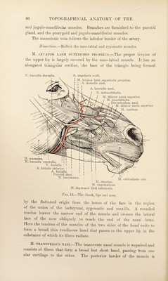
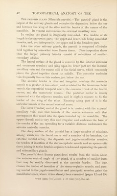
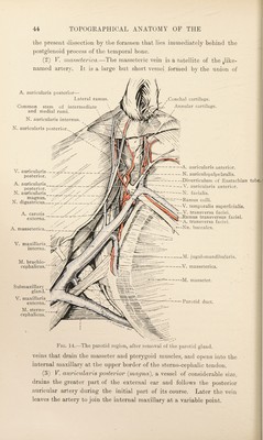
![The parotid gland (Glandula parotis).—The parotid ^ gland is the largest of the salivary glands and occupies the depression below the ear and between the wing of the atlas and the border of the ramus of the mandible. Its ventral end reaches the external maxillary vein. In outline the gland is irregularly four-sided. The middle of its length is the narrowest part; the upper and lower ends being somewhat broader, and, not infrequently, the upper end is the broadest part. Like the other salivary glands, the parotid is composed of lobules held together by somewhat loose fibrous tissue. Close inspection shows that the larger, primary lobules consist of collections of smaller, secondary lobules. The lateral surface of the gland is covered by the inferior auricular and cutaneous muscles ; and lying upon its lower part are the internal maxillary vein and the ramus colli of the facial nerve, which commonly pierce the gland together about its middle. The posterior auricular vein frequently lies on this surface just below the ear. The anterior border is thin and irregular, overlaps the masseter muscle to a greater or less extent, and is related to the transverse facial vessels, the superficial temporal nerve, the common trunk of the buccal nerves, and the masseteric vessels. The posterior border is loosely connected with the adjacent muscles, and is slightly concave to fit the border of the wing of the atlas. Running along part of it is the auricular branch of the second cervical nerve. The lower (ventral) end of the gland is in contact with the external maxillary vein, and that branch of the nervus cutaneus colli that accompanies this vessel into the space bounded by the mandible. The upper (dorsal) end is very thin and irregular, and embraces the base of the concha of the ear, spreading for a variable distance underneath the anterior auricular muscles. The deep surface of the parotid has a large number of relations, among which are the facial nerve and a number of its branches, the external carotid artery, the digastric and jugulo-mandibular muscles, the tendon of insertion of the sterno-cephalic muscle and an aponeurotic sheet joining it to the brachio-cephalic tendon and separating the parotid and submaxillary glands. The 'parotid duct (ductus parotideus) results from the union, about the anterior ventral angle of the gland, of a number of smaller ducts that may be readily discovered at the anterior border. The duct crosses the tendon of insertion of the sterno-cephalic muscle, and, pass¬ ing medial to the jugulo-mandibular and pterygoid muscles, gains the mandibular space, where it has already been examined (pages 12 and 39). 1 Trapd (para) [Gr.], about + ods (ous) [Gr.], the ear.](https://iiif.wellcomecollection.org/image/b29820066_0058.jp2/full/800%2C/0/default.jpg)