The topographical anatomy of the head and neck of the horse / [O. Charnock Bradley].
- Orlando Charnock Bradley
- Date:
- 1923
Licence: In copyright
Credit: The topographical anatomy of the head and neck of the horse / [O. Charnock Bradley]. Source: Wellcome Collection.
78/248 page 62
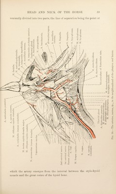
![vessel that follows the auditory (Eustachian) tube and gains the middle ear by the petro-tympanic fissure. (4) A. meningea media.—The middle meningeal artery is also small and arises from the internal maxillary as it is approaching the entrance to the alar canal. It at once gains the cranium by the foramen spinosum. (5) A. temporalis profunda posterior.—The posterior deep temporal artery arises at the entrance to the alar canal and thence passes upwards and backwards underneath the temporal muscle. It anastomoses with the middle meningeal and superficial temporal arteries, and supplies a branch to the masseter muscle. N. MA.ND1BULARIS.—The fifth cerebral, or trigeminal, nerve divides into three parts while still within the cranium, and each part leaves the cranium by a different foramen. The three nerves formed by the trigeminal are the ophthalmic, the maxillary and the mandibular ; and it is the last-named that can be examined at the present time. In view of its distribution, it should be remembered that the mandibular ^ nerve differs from the ophthalmic and the maxillary in that, though mainly composed of sensory fibres, it contains motor fibres as well. The mandibular nerve is of large size and leaves the cranium by the foramen ovale. Immediately on its exit from the foramen it divides into a number of branches of varying size. (1) N. massetericus.—The masseteric nerve curves round the front of the mandibular joint in company with a tributary of the transverse facial vein, and traverses the mandibular notch to reach the masseter muscle. The terminal part of its course was seen during the removal of the masseter. (2) Nn. temporales profundi.—The deep temporal nerves are generally two in number, and usually arise from the mandibular in common with the masseteric nerve. They pass upwards and forwards to enter the temporalis muscle. (3) N. pterygoidetis.—The pterygoid nerve is the smallest branch of the mandibular. It supplies the pterygoid muscles. A small, flat, oval ganglion—the otic - ganglion (ganglion oticum)— is placed at the origin of the pterygoid nerve, where it rests upon the tensor of the soft palate. The ganglion receives motor fibres from the pterygoid nerve, sensory fibres from the facial and glosso-pharyngeal nerves by way of the lesser superficial petrosal nerve, and sympathetic filaments from the plexus about the internal maxillary artery. Delicate nerves pass from the ganglion to the tensor tympani, the tensor of the soft palate and the auditory tube. ^ Mandihulum [L.], the lower jaw. 2 wrt/co's (oticos) [Gr.], pertaining to the ear (oi’s).](https://iiif.wellcomecollection.org/image/b29820066_0078.jp2/full/800%2C/0/default.jpg)