The topographical anatomy of the head and neck of the horse / [O. Charnock Bradley].
- Orlando Charnock Bradley
- Date:
- 1923
Licence: In copyright
Credit: The topographical anatomy of the head and neck of the horse / [O. Charnock Bradley]. Source: Wellcome Collection.
86/248 page 70
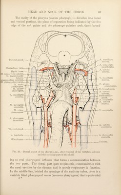
![into a gap between the pharyngeal muscles, and is, consequently, in direct contact with the diverticulum of the auditory tube. The ventral part (pars digestoria) is both respiratory and alimentary in function, inasmuch as it communicates with the mouth and dorsal part in the one direction and with the oesophagus and larynx in the other. On each side of the entrance to the larynx there is a narrow but deep 'piriform recest^ (recessus piriformis). The mucous membrane of the upper part of the cavity is redder than that of the more ventral and caudal part. There are seven openings into the cavity of the pharynx. Of these the opening from the mouth (isthmus faucium), the opening into the larynx (aditus laryngis) and the continuation of the cavity into the oesophagus, are single and median, and occur in the pars digestoria. Two paired openings, namely, from the nasal cavities (choanse) and from the auditory tubes, are present in the dorsal part. The openings of the auditory (Eustachian) tubes, the ostia'^ pharyngea tuhcB, are slit-like apertures, covered medially by the flattened terminations of the cartilaginous wall of the tubes. A small oblique fold of mucous membrane (plica salpingopharyngea) runs down¬ wards from each opening towards the larynx. Dissection.—The small opening that was previously made in the wall of the diverticulum of the auditory tube should be extended by a hori¬ zontal incision sufficiently large to show the whole of the interior of the cavity. Make the incision half-way between the stylo-pharyngeal muscle and the auditory tube. The auditory (eustachian) ^ tube and its diverticulum— Diverticulum tuboe auditivce.—The diverticulum of the auditorv tube •/ is a thin-walled sac, capable of holding some 300 cc. of fluid, formed by the extrusion of the mucous membrane of the tube out of an elongated slit in its ventral wall. Its occurrence, among domesticated mammals, is peculiar to the Equidce. The diverticulum occupies the whole of the space between the base of the cranium, the atlas and the pharynx; and (except where the ventral straight muscles of the head intervene) it comes into contact with its fellow in the median plane, where a thin partition, formed by the fusion of the mucous linings of the two diverticula, is all that separ¬ ates the interior of the two cavities. The anterior limit of the cavity is in the form of a small blind pouch immediately ventral to the body of the anterior part of the sphenoid bone and a short distance from the choanse; or, expressed in terms of 1 Ostium (dim. of os, a mouth) [L.], a small opening. ^ Bartolomeo Eustachio, an Italian anatomist, 1520-1574.](https://iiif.wellcomecollection.org/image/b29820066_0086.jp2/full/800%2C/0/default.jpg)