The topographical anatomy of the head and neck of the horse / [O. Charnock Bradley].
- Orlando Charnock Bradley
- Date:
- 1923
Licence: In copyright
Credit: The topographical anatomy of the head and neck of the horse / [O. Charnock Bradley]. Source: Wellcome Collection.
93/248 page 77
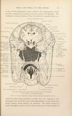
![layers:—(1) The relatively thick oral mucous membrane continuous with that of the hard palate; (2) a layer of [Hilatine ;jh(n<h; (d) an aponeurotic and muscular layer ; and (4) the pharyngeal mucous membrane continuous with that of the nasal cavity. DUi^ectvni.—Dissect the mucous membrane from the pharyngeal surface of the soft ])alate. The muscular and aponeurotic layer contains the origin of the palato-pharyngeal muscle (already dissected in connection with the wall of the ])harynx), the termination of the levator and tensor muscles of the palate, and the muscle of the uvula (m. uvuhe). It is convenient to dissect the whole length (d the tensor and levator muscles at this stage. M. UVUL.I*:.—The muscle of the uvula^ is often described as being unpaired, but probably the best way to regard it is as a pair of muscles (right and left) blended in the middle line of the soft palate. It has a wide aponeurotic origin from the free border of the palatine bone, and ends near the free border of the soft palate. A[. TENSOR VELI PALATINI.—The tensor of the soft palate lies along the lateral surface of the auditory tube. Arising from the muscular process of the temporal bone, the lateral lamina of the cartilage of the auditory \ tube and the pterygoid bone, the muscle ends in a narrow, flattened tendon that bends round the hamulus of the pterygoid bone to end in the general aponeurosis of the soft palate. A small synovial bursa facilitates the play of the tendon round the pterygoid hamulus. M. LEVATOR VELI PALATINI.—The levator of the soft palate arises from the muscular process of the temporal bone and the lateral lamina of the cartilage of the auditory tube in common with the tensor, medial to which it lies. The two levator muscles (right and left) enter the substance of the soft palate close together. Dissection.—Reflect the muscular and aponeurotic layer of the soft palate so as to expose the glands. The greyish-yellow pahitine (jlands (glandula* palatinse) form a layer of 1 cm. or more in thickness. It is important to note that their ducts open only on the ventral or oral surface of the soft palate; that is, the mucous glandular secretion is poured out on that surface with which the food comes into contact during its passage from the mouth to the pharynx. Dissection.—Make a longitudinal incision through the most lateral part of what still nmiains of the soft palate. The mouth (Cavum oris).—The cavity of the mouth is the initial part of the alimentary tract and extends from the lips to the isthmus ^ Uvuhi [L.], dim. of ui'a, a grape.](https://iiif.wellcomecollection.org/image/b29820066_0093.jp2/full/800%2C/0/default.jpg)