Paediatric implications for some adult disorders : Scientific proceedings of the 4th Unigate Workshop, held at the Royal Academy of Physicians, St. Andrews Place, London, N.W. 1, May 1976 / edited by Donald Barltrop.
- Unigate Paediatric Workshop 1976 : Royal College of Physicians)
- Date:
- 1977
Licence: Attribution-NonCommercial-NoDerivatives 4.0 International (CC BY-NC-ND 4.0)
Credit: Paediatric implications for some adult disorders : Scientific proceedings of the 4th Unigate Workshop, held at the Royal Academy of Physicians, St. Andrews Place, London, N.W. 1, May 1976 / edited by Donald Barltrop. Source: Wellcome Collection.
29/168 page 17
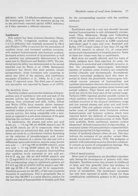
![Metabolic causes of renal stone formation 17 deficiency with 2,8-dihydroxyadeninuria represent the homozygous state for the mutation giving rise to the previously reported partial APRT deficiency or if they represent a difl'erent mutation. Xanthinuria This subject has been reviewed elsewhere (Watts, 1976c; 1977b). Congenital xanthine oxidase (EC 1.2.3.2) deficiency was originally proposed by Dent and Philpott (1954) to account for the association of xanthine stone, and increased xanthine excretion, with marked hypouricaemia and hypouric aciduria. Increased urinary excretion of hypoxanthine as well as xanthine was subsequently demonstrated in the same case by Dickinson and Smelile (1959). The pre¬ dicted enzyme defect was demonstrated in the second reported case by Watts et al. (1964). Subsequent experience has shown that most patients remain asymptomatic, stone formation only occurring in about one third of the patients, and xanthinuric myopathy (Chalmers et ah, 1969a, b) in 3 out of about 25 reported cases. The third case of xanthin¬ uric myopathy was reported by Isaacs et al. (1975). The metabolic lesion Xanthine oxidase catalyses the oxidation of hypox¬ anthine and of xanthine to uric acid and lack of the enzyme activity has been demonstrated in jejunal mucosa, liver, colostrum and milk. Gibbs, Allsop and Watts (1976) have recently shown immuno- chemically that a xanthinuric patient's milk lacks xanthine oxidase protein as well as xanthine oxidase enzyme activity, this being compatible with a non¬ sense as opposed to a mis-sense mutation of the gene directing the synthesis of xanthine oxidase. In xanthinuria, the plasma and urine uric acid levels are <10 mg/100 ml (0-0596 mmol/1) and 50 mg (0-295 mmol) per 24 hr respectively when the patient takes an unrestricted diet, and the measure¬ ments are made by a colorimetrie method depending on the reduction of phosphotungstic acid. Lower results are obtained on a purine free diet and with a diff'erential spectrophotometric enzyme assay [serum uric acid = < 0-5 mg/100 ml (0-0298 mmol/1), urine uric acid <15 mg (0-089 mmol) per 24 hr]. The normal plasma and urine oxypurine (hypoxanthine plus xanthine) levels expressed as uric acid are: 0-0-0-25 mg per 100 ml (0-0-0-0149 mmol/1) and 11-22 mg (0-0655-0-131 mmol) per 24 hr respectively. The corresponding values in xanthinuric patients are 0-5-0-9 mg/100 ml (0-0295-0-0536 mmol/1) and 100- 600 mg (0-596-3-58 mmol) per 24 hr respectively. Xanthine accounts for 60-90% of the total oxypurine excretion in xanthinuria. This is presumably due to the fact that HGPRT is a more effective catalyst for the conversion of hypoxanthine to inosinic acid than for the corresponding reaction with the xanthine substrate. Genetics Xanthinuria must be a very rare disorder because marked hypouricaemia is only infrequently encoun¬ tered. Thus, Mikkelson, Dodge and Valkenberg (1965) found no serum uric acid values of less than 1-0 mg/100 ml (0-059 mmol/1) in 6,000 unselected individuals aged 4 years and over. Ramsdell and Kelley (1973) found values of less than 2-0 mg/100 ml (0-118 mmol/1) in almost 1% of consecutive serum urate measurements in hospital practice. None of these low values were due to xanthinuria. Both sexes are equally affected, and two types of family pedigree have been reported. In some, the inheritance is autosomal and completely recessive, in that the presumably heterozygous individuals (parents of xanthine stone formers) are completely normal clinically and biochemically. Incompletely recessive autosomal pedigrees have also been re¬ ported in which the presumably heterozygous indi¬ viduals excrete amounts of hypoxanthine and xanthine intermediate between those excreted by the presumably homozygous xanthine stone formers and normal subjects. Their blood and urine uric acid levels are also in the lower part of the normal range. Terhorst (1969) reported another possible variant in which xanthine stone formation, hypoxanthine and xanthine excretion in the classical xanthinuric range and low normal plasma and urine uric acid levels co-existed. This case appears similar to some of the earlier cases of xanthine stone formation without hypouricaemia and hypouric aciduria which were reported before the methods of measuring hypox¬ anthine and xanthine concentrations in body fluids were available. Some xanthinuric patients oxidize allopurinol [4-hydroxypyrazolo(3,4-i/)pyrimidine, a structural isomer of hypoxanthine] to oxipurinol [4,6- dihydroxypyrazolo(3,4-ii)pyrimidine, a structural isomer of xanthine] whereas others do not. This may indicate the existence of yet another variant (Sim- monds. Levin and Cameron, 1974). The pedigrees of patients with autosomal recessive disorders common¬ ly contain an excess of consanguinous matings. This has not so far been reported in xanthinuria. Clinical manifestations Most casts of xanthinuria are identified coinciden- tally during the investigation of patients with other disorders. Xanthine stones may present in childhood or later, and they generally show less tendency to recur than other stones which are due to inborn errors of metabolism. The xanthinuric myopathy has only been reported in adults. Except for these complications xanthinuria appears to be a harmless trait.](https://iiif.wellcomecollection.org/image/b18031298_0030.JP2/full/800%2C/0/default.jpg)