Paediatric implications for some adult disorders : Scientific proceedings of the 4th Unigate Workshop, held at the Royal Academy of Physicians, St. Andrews Place, London, N.W. 1, May 1976 / edited by Donald Barltrop.
- Unigate Paediatric Workshop 1976 : Royal College of Physicians)
- Date:
- 1977
Licence: Attribution-NonCommercial-NoDerivatives 4.0 International (CC BY-NC-ND 4.0)
Credit: Paediatric implications for some adult disorders : Scientific proceedings of the 4th Unigate Workshop, held at the Royal Academy of Physicians, St. Andrews Place, London, N.W. 1, May 1976 / edited by Donald Barltrop. Source: Wellcome Collection.
30/168 page 18
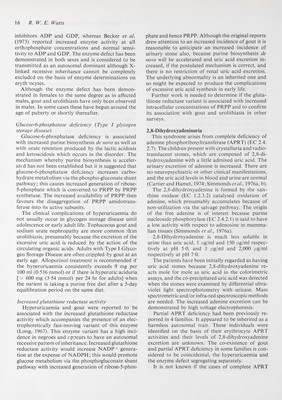
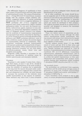
![18 R.W. E. Watts The differential diagnosis of xanthinuria is from other causes of hypouricaemia and from other causes of radiotranslucent stones. The causes of hypouricae¬ mia [< 2'0 mg/100 ml (0-118 mmol/1)] are: excessive dosage with the xanthine oxidase inhibitor allo- purinol, congenital deficiency of purine nucleoside Phosphorylase (EC 2.4.2.1), congenital deficiency of PRPP synthetase (EC 2.7.6.1), congenital and acquired renal tubule reabsorption defects (isolated reabsorption defect for uric acid, the Fanconi syn¬ drome, radiocontrast agents, exposure to toxic sub¬ stances, e.g. tetracycline degradation products), rare cases of malignant disease, extensive liver disease, and the syndrome of hypouricaemia, hypercalciuria and decreased bone density described by Sperling et al. ( 1974). Except for purine nucleoside Phosphorylase deficiency and PRPP synthetase deficiency, none of these conditions usually produce the extreme degree of hypouricaemia observed in xanthinuria. Only, allopurinol overdosage produces hypouricaemia with hypouric aciduria and increased levels of hypox- anthine and xanthine. Other radiotranslucent lesions causing obstructive uropathy are uric acid stones, 2,8-dihydroxyadenine stones, blood clots and sloughed renal papillae. Seegmiller (1968) describes a procedure for the unequivocal identification of xanthine stones by modern spectroscopic and enzymatic methods. Treatment Treatment is only needed if stones form, when a high fluid intake, at least 3 1 per 24 hr, should be recommended. Alkalinization of the urine would in¬ crease the solubility of xanthine but is undesirable in the long term for reasons discussed under cystin- uria. Some authors have suggested that a purine free diet and allopurinol are valuable, the latter because it increases the excretion of the relatively more soluble hypoxanthine at the expense of xanthine. These measures may merit trial if stone formation persists in spite of an adequate water diuresis and alkalinization of the urine. As in other conditions, the stones should be re¬ moved surgically only if : (i) they are causing urinary obstruction and will not pass spontaneously, (ii) their presence appears to be predisposing to recurrent urinary tract infection; (iii) they are causing pain which can clearly be attributed to their presence. The surgical procedure should be as conservative as possible, and designed to preserve the maximum amount of renal tissue. The hereditary orotic acidurias These two inborn errors of metabolism are in¬ cluded in this review because although the other clinical manifestations usually predominate, they may present with urinary tract obstruction by masses of orotic acid crystals. The crystals do not usually pack together to form stones. Normal subjects excrete about 1-5 mg (0-009 mmol) of orotic acid per 24 hr in their urine, and this is greatly increased in the hereditary orotic acid¬ urias. Smaller increases are observed in: Type II hereditary hyperammonaemia [ornithine carbamoyl- transferase (EC 2.1.3.3) deficiency]; 6-azauridine administration; allopurinol administration; and in the hereditary orotic aciduria variant with resistance to uridine but partial responsiveness to folic acid. Allopurinol and 6-azauridine both produce orotic aciduria because their metabolites inhibit orotidine 5'-phosphate (OMP) decarboxylase (EC 4.1.1.23). The metabolic lesions The pyrimidines are synthesized from aspartate, bicarbonate and the amido group nitrogen atoms of glutamine via orotic acid, orotidylic acid [orotidine 5'-phosphate (OMP)] and uridylic acid [uridine 5'- phosphate (UMP)] (Fig. 3). Type I hereditary orotic aciduria is due to defi¬ ciency of orotate phosphoribosyltransferase (OPRT) CARBAMYL PHOSPHATE DIHYDRO-OROTATE CARBAMYL Dihydro-orotase Carbamyl transferase ASPARTATE (EC 3.5.2.3) (EC2.1.3.2} Dihydro-orotate oxidase EC 1.3.3.1 Uridine Kinase OMP-decarboxylase Orotate URIDINE ►►UMP'* ] ( OMP^- ) [ OROTATE (EC 2.7.1.48) phosphoribosyl transferase (EC 2.4.2.10) Nucleoside- monophosphate kinase (EC 2.7.4.4) NUCLEIC ACIDS Fig. 3. Pyrimidine biosynthesis. The sites of the metabolic lesions in the hereditary orotic acidurias are shown by broken arrows.](https://iiif.wellcomecollection.org/image/b18031298_0031.JP2/full/800%2C/0/default.jpg)