Plagiostomata of the Pacific. Pt. I. Fam. Heterodontidae / by N. de Miklouho-Maclay and William Macleay.
- Nicholas Miklouho-Maclay
- Date:
- [1878]
Licence: Public Domain Mark
Credit: Plagiostomata of the Pacific. Pt. I. Fam. Heterodontidae / by N. de Miklouho-Maclay and William Macleay. Source: Wellcome Collection.
Provider: This material has been provided by The Royal College of Surgeons of England. The original may be consulted at The Royal College of Surgeons of England.
28/34
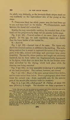
![the adult, very distinctly, as the brownish-black stripes stood out very markedly on the light-colored skin of the young at this stage. #—Transverse black bar which passes over the head from eye to eye, and loses itself on the cheeks. **—Characteristic mark between the dorsal and ventral fins. Besides the very remarkable marking, the rounded form of the head and the proportionally large tail are peculiar to this stage. Fig. 2 (pi. 22).—Ventral surface of the same (from a photo- graph). At this age, the male copulatory organs are shorter than the lower border of the ventral fin. fa.—Anal fin. Fig. 5 (pi. 23).—Lateral view of the same. The figure only shews the external contour, in addition to the marking. The undu- lating contour-line is meant to represent the extent of the rougher parts of the skin, covered with large and prominent bony plates (scutellce). The anal fin, whose position and length are accurately rendered, has its form rather too diagrammatically represented in the figure, which does not shew that the fin has become some- what shrivelled by the drying, which took place while the drawing was being executed. Fig. 6 (pi. 23).—A very miserable rendering of a photograph of the head frombefore. The outlines, however, are correct. Fig. 7. (pi. 23).—Head of the same animal viewed from before, and to some extent from below. From a photograph, about three times the natural size. Scale applicable only for the anterior part of the mouth (the anterior teeth for instance). Figs. 3, 4, 8 (pi. 23).—Full-grown* * H. Phillipi, of about 795 mm. (31'4 in.) in length. The sketches are from a specimen in * N.B.—The scale can only be relied on for a certain part of the object (the part on the same plane with it), and must, therefore, be used with caution. * The largest specimen I have seen in Sydney was a female of about 1,232 mm. (48'5 in.) in length. As the specimen was a dried and stuffed one (by which means the shape of the head is considerably altered), its length in the fresh state w'as probably greater. From the external point of the one fin to that of the other it measured 602 mm. (which number also is to be regarded as only approximate). t I received the first fresh specimen of II. Phillipi on the twentieth day after my arrival in Sydney; but could make drawings much earlier from specimens preserved in in spirit in the Macleay Museum. As the proportions of the different parts of the body as well as the form of the fins are not much altered by the action of alcohol for a moderate time on the specimen, I found it unnecessary to waste time in making any fresh drawings. [26]](https://iiif.wellcomecollection.org/image/b22367913_0028.jp2/full/800%2C/0/default.jpg)