Plagiostomata of the Pacific. Pt. I. Fam. Heterodontidae / by N. de Miklouho-Maclay and William Macleay.
- Nicholas Miklouho-Maclay
- Date:
- [1878]
Licence: Public Domain Mark
Credit: Plagiostomata of the Pacific. Pt. I. Fam. Heterodontidae / by N. de Miklouho-Maclay and William Macleay. Source: Wellcome Collection.
Provider: This material has been provided by The Royal College of Surgeons of England. The original may be consulted at The Royal College of Surgeons of England.
30/34
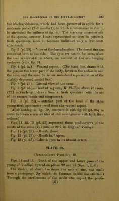
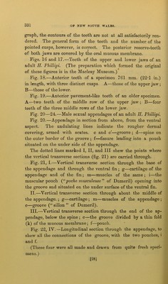
![graph, the contours of the teeth are not at all satisfactorily ren- dered. The general form of the teeth and the number of the pointed cusps, however, is correct. The posterior reserve-teeth of both jaws are covered by the oral mucous membrane. Figs. 16 and 17.—Teeth of the upper and lower jaws of an adult H. PJiillipi. (The preparation which formed the original of these figures is in the Macleay Museum.) Fig. 18.—Anterior teeth of a specimen 761 mm. (22T in.) in length, with three distinct cusps. A—those of the upper jaw ; B—those of the lower. Fig. 19.—Anterior pavement-like teeth of an older specimen. A—two teeth of the middle row of the upper jaw ; B—four teeth of the three middle rows of the lower jaw. Fig. 20—24.—Male sexual appendages of an adult E. Phillipi. Fig. 20.—Appendage in section from above, from the ventral aspect. The undulating lines indicate the rougher dermal covering, armed with scutes, c and c'—groove ; d—spine on the outer border of the groove ; f—fissure leading into a pouch situated on the under side of the appendage. The dotted lines marked I, II, and III shew the points where the vertical transverse sections (fig. 21) are carried through. Fig. 21, I.—Vertical transverse section through the base of the appendage and through the ventral fin ; g—cartilage of the appendage and of the fin; m—muscles of the same ; i—the muscular pouch (“poche musculeuse” of Dumeril) opening into the groove and situated on the under surface of the ventral fin. II. —Vertical transverse section through about the middle of the appendage. ; g—cartilage ; m—muscles of the appendage ; c—groove (“ sillon ” of Dumeril). III. —Vertical transverse section through the end of the ap- pendage, below the spine ; c—the groove divided by a thin fold (k) of the mucous membrane; f—pouch. Fig. 22, IV. —Longitudinal section through the appendage, to shew all the connections of the groove, with the two pouches, i and f. (These four were all made and drawn from quite fresh speci- mens.) [28]](https://iiif.wellcomecollection.org/image/b22367913_0030.jp2/full/800%2C/0/default.jpg)