The essentials of histology, descriptive and practical, for the use of students.
- Edward Albert Sharpey-Schäfer
- Date:
- 1914
Licence: In copyright
Credit: The essentials of histology, descriptive and practical, for the use of students. Source: Wellcome Collection.
Provider: This material has been provided by Royal College of Physicians, London. The original may be consulted at Royal College of Physicians, London.
410/436 page 394
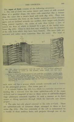
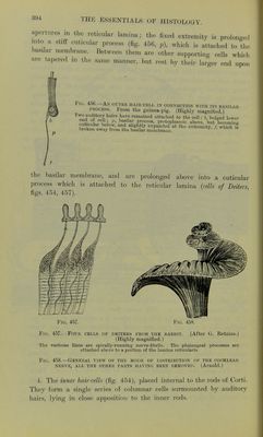
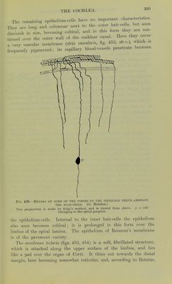
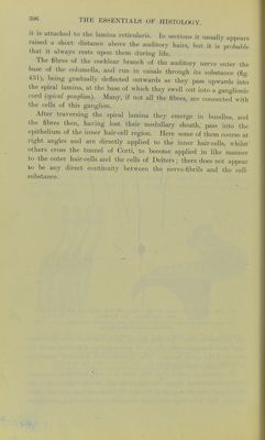
![apertures in the reticular lamina j the fixed extremity is prolonged into a stiff cuticular process (fig. 45G, p), which is attached to the basilar membrane. Between them are other supporting cells which are tapered ,n the same manner, but rest by their larger end upon Fig. 4oC—An outer hair-cell in connection with its basilar process. From the guinea-pig. (Highly magnified.) 3U51™71 !mirfl ]»lv.f remained attached to the cell; 6, bulged lower ena ol cell v, basilar process, protoplasmic above, but becoming ffiS™ b0l°fW' alf e^anded at the extremity /, whiS if broken away from the basilar membrane. the basilar membrane, and are prolonged above into a cuticular process which is attached to the reticular lamina (cells of Deiters, figs. 454, 457). Fig. 457. Fig. 458. Fis. 457.- Four cells of deiters from the rabbit. (After G. Retzius.) (Highly magnified.) The varicose lines are spirally-running nerve-fibrils. The phalangeal processes are attached above to a portion of the lamina reticularis. Fie 458.—General view of the mode of distribution of the cochlear nerve, all the other parts having been removed. (Arnold.) 4. The inner hair-celh (fig. 454), placed internal to the rods of Corti. They form a single series of columnar cells surmounted by auditory hairs, lying in close apposition to the inner rods.](https://iiif.wellcomecollection.org/image/b24758474_0410.jp2/full/800%2C/0/default.jpg)