A treatise on the diseases of the ear including the anatomy and physiology of the organ together with the treatment of the affections of the nose and pharynx which conduce to aural disease.
- Hovell, Mark, 1853-1925
- Date:
- 1894
Licence: Public Domain Mark
Credit: A treatise on the diseases of the ear including the anatomy and physiology of the organ together with the treatment of the affections of the nose and pharynx which conduce to aural disease. Source: Wellcome Collection.
Provider: This material has been provided by the Harvey Cushing/John Hay Whitney Medical Library at Yale University, through the Medical Heritage Library. The original may be consulted at the Harvey Cushing/John Hay Whitney Medical Library at Yale University.
42/752
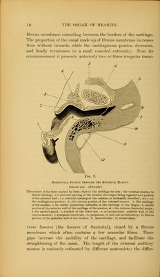
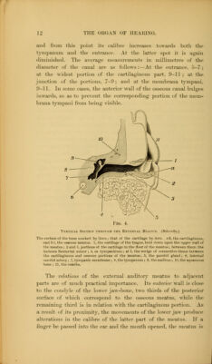
![On tlie ])osterior surface of the petrous portion is the narrow fissure for the aquwdttdiis vestihuli. This is covered by a depressed scale of bone, and is situated three lines behind the internal auditory meatus. Through this aperture purulent secretion some- times passes into the cranial cavity from the tympanum and vestibule. Another small aperture, for the afpuedudiis cocldew, begins in a triangular wider depression in the posterior border •directly below the internal auditoiy meatus. This canal passes up- wards and outwards through the substance of the petrous bone to the scala tiimpani. and through it a communication takes place between the latter and the subarachnoid space. At the base of the skull, in the plate of bone between the jugular fossa and the carotid ■canal, is a small foramen through which the nerve of Jacohson •(from the petrous ganglion of the glosso-pharyngeal) passes to the tympanum, and in the wall of the carotid canal is a small opening for the tt/mjKinic h'anch of the carotid plexus. The auricular branch of the vagus passes in a groove and foramen in the jugular fossa, and close to the canal for the tensor tympani muscle is a foramen for the small superficial petrosal nerve. The lanfe nerve of that name, from Meckel's ganglion, through the Vidian nerve, passes in a groove on the anterior surface of the petrous portion to a foramen, the hiatus Falloptii, and is thus conducted to the -aqueduct, where it joins the gangliform enlargement of the facial nerve. The integument of the meatus is continuous with that of the Auricle, but gradually becomes thinner and more delicate. It is firmly attached to the wall of the canal, and at the inner extremity it is stretched over the membrana tympani, of which it forms the •external layer. It is easily detached from that structure after maceration in water, or when decomposition has occurred; the •cuticular lining of the canal can then be drawn out in the form of ■a tube closed at one end. The skin of the external meatus exhibits certain peculiarities in •different parts. In the cartilaginous portion and on the roof of that part of the osseous meatus which is formed by the squamous bone, it resembles the ordinary integument. But in the remaining larger portion of the osseous meatus, it is very thin (not more than one-tenth of a millimetre in thickness), and closely blended with the periosteum, so as to form a shining fibrous membrane. Neither liairs nor glands are present; but near the membrana tympani](https://iiif.wellcomecollection.org/image/b21019423_0042.jp2/full/800%2C/0/default.jpg)