Diseases of the heart / By John Cowan with chapters on the electro-cardiograph, by W.T. Ritchie and the ocular manifestations in arterio-sclerosis, by Arthur J. Ballantyne.
- Cowan, John, 1870-
- Date:
- 1914
Licence: Public Domain Mark
Credit: Diseases of the heart / By John Cowan with chapters on the electro-cardiograph, by W.T. Ritchie and the ocular manifestations in arterio-sclerosis, by Arthur J. Ballantyne. Source: Wellcome Collection.
Provider: This material has been provided by the Augustus C. Long Health Sciences Library at Columbia University and Columbia University Libraries/Information Services, through the Medical Heritage Library. The original may be consulted at the the Augustus C. Long Health Sciences Library at Columbia University and Columbia University.
25/484
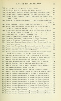
![FIG. PAGE 141. Cheonic Mitral and Atjeiculae Endocarditis . . . 266 142. Syphilitic Disease of Aortic and Mitral Valves - - 268 143. Chronic Renal Disease: Patchy Thickening of Mitral Cusps - 272 144. Chronic Renal Disease: Diffiise Thickening of Mitral Cusps - 273 145. Chronic Renal Disease: Diffuse Thickening of Aortic and Mitral Cusps ....... 274 146. Brachial and Respiratory Curves in Cheyne-Stokes Breathing facing 287 147. Blood-Pressure Tracing: Aortic Regurgitation - - - 300 148. DlAGRAJil SHO\VING SURFACE RELATIONS OF THE LuNGS, HeART, AND Cardiac Valves (A. Keith) - - - - - 321 149. Diagram shoaving Surface Relations of the Pericardium, Heart, AND Great Vessels (A. Keith) ..... 322 150. Curves showing In-verted Apex-Impulse - - - 323 151. Curves of Epigastric Pulsation due to Aoria - - - 323 152. Curves of Epigastric Pulsation due to Right Ventricle - 323 153. Curves of Epigastric Pulsation due to Left Ventricle - - 324 154. Curves from Brachial Artery, Second Left Intercostal Space, AND Apex-Impulse ....... 324 155. Curves from Fourth Right Intercostal Space and Apex-Impulse 324 156. Diagraji showing Surface Relations of Heart and Lungs - 325 157. DiAGRAii showing Area of Cardiac Dulness - - - 326 158. Diagram showing Time Relations of the Cardiac Contractions, THE Cardiac Sounds, and the Valvular Murmurs - - 328 159. Skiagram: Hypertrophy of Left Ventricle (J. R. Riddell) - 334 160. Diagram showing Area of Cardiac Dulness in Mitral Stenosis - 335 161. Diagram showing Distribution of Presystolic Murmur - - 337 162. Skiagram: Hypertrophy of Right Ventricle (J. R. Riddell) - 338 163. Diagram showing Area of Reduplicated Second Sound - - 340 164. Diagram showing Area of Early Diastolic Mitral Murmur - 340 165. Blood-Pressure Chart: Mitral Disease - . . . 341 166. Blood-Pressure Chart: Mitral Disease; Cirrhotic Kidney - 342 167. Diagram showing Area of Cardiac Dulness in a Dilated Heart 343 168. Diagram showing Area of Cardiac Dulness in Mitral Reflux - 343 169. Diagram showing Distribution op Murmur in Mitral Reflux - 344 170. Diagram showing Distribution of Murmur in Mitral Reflux - 344 171. Diagram showing Distribution of Murmur in Aortic Stenosis - 348 172. Diagram showing Distribution of Murmur in Aortic Stenosis - 348 173. Diagram showing Area of Cardiac Dulness in Aortic Reflux - 349 174. Diagram showing Distribution of Murmur in Aortic Reflux - 350 175. Diagram showing Distribution of Murmur in Aortic Reflux - 350 176. Sphygmogram: Aortic Reflux ..... 351 177. Diagram showing Distribution of Murmur in Pulmonary Stenosis 355 178. Diagram showing Distribution of ]\1urmur in Pulmonary Reflux - 355](https://iiif.wellcomecollection.org/image/b2122531x_0025.jp2/full/800%2C/0/default.jpg)