A system of human anatomy, general and special / By Erasmus Wilson.
- William James Erasmus Wilson
- Date:
- 1843
Licence: Public Domain Mark
Credit: A system of human anatomy, general and special / By Erasmus Wilson. Source: Wellcome Collection.
Provider: This material has been provided by the Augustus C. Long Health Sciences Library at Columbia University and Columbia University Libraries/Information Services, through the Medical Heritage Library. The original may be consulted at the the Augustus C. Long Health Sciences Library at Columbia University and Columbia University.
561/596 page 547
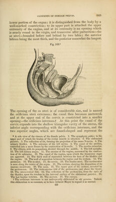
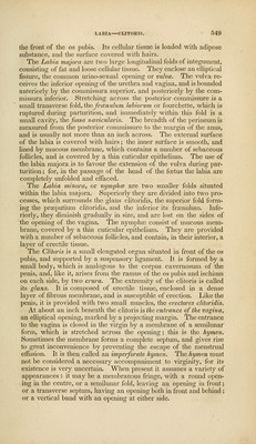
![The Nerves are derived from the hypogastric and spermatic plexuses, and from the sacral plexus. The Jlppendages of the uterus are enclosed by the lateral dupli- catures of peritoneum, called the broad ligaments. They are the Fallopian tubes and ovaries. FALLOPIAJf TUBES. The Fallopian* tubes or oviducts, the uterine trumpets of the French writers, are situated in the upper border of the broad liga- ments, and are connected with the superior angles of the uterus. They are somewhat trumpet-shaped, being much smaller at the uterine than at the free extremity, and narrower in the middle than at either end. Each tube is about four or five inches in length, and more or less flexuous in its course. The canal of the Fallopian tube is exceedingly minute, its inner extremity opens by means of the ostium uterinum into the upper angle of the cavity of the uterus, and the opposite end into the cavity of the peritoneum. The free or expanded extremity of the Fallopian tube presents a double and sometimes a triple series of small processes or fringes which sur- round the margin of the trumpet or funnel-shaped opening, the ostium abdominale. This fringe-like appendage to the end of the tube has gained for it the appellation of the fimbriated extremity; and the remarkable manner in which this circular fringe applies itself to the surface of the ovary during sexual excitement, the additional title of morsus diaboli. One of these processes longer than the rest, or, according to Cruveilhier a distinct ligamentous cord, is attached to the distal end of the ovary, and serves to guide the tube in its seizure of that organ. The Fallopian tube is composed of three tunics, an external and loose investment derived from the peritoneum; a middle or muscular coat consisting of circular [internal] and longitudinal [external] fibres, continuous with those of the uterus; and an internal or lining mucous membrane which is continuous on the one hand with the mucous membrane of the uterus, and at the opposite extremity with the peritoneum. In the minute canal of the tube the mucous membrane is thrown into longitudinal folds or rugas, which indicate the adaptation of the tube to dilatation. OVARIES. The Ovaries are two oblong flattened and oval bodies of a whitish colour, situated in the posterior layer of peritoneum of the broad ligaments. They are connected to the upper angles of the uterus * Gabriel Fallopius, a nobleman of Modcna, was one of the founders of modern anatomy. He was Professor at Ferrara, then at Pisa, and afterwards succeeded Vesa- lius at Padua. His principal observations are collected in a work, Observationes Anatomicoe, which he published in 1561.](https://iiif.wellcomecollection.org/image/b21203933_0561.jp2/full/800%2C/0/default.jpg)
 No text description is available for this image
 No text description is available for this image
 No text description is available for this image