The skeleton in the flying lemurs, Galeopteridae / by R.W. Shufeldt.
- Robert Wilson Shufeldt
- Date:
- 1911
Licence: In copyright
Credit: The skeleton in the flying lemurs, Galeopteridae / by R.W. Shufeldt. Source: Wellcome Collection.
Provider: This material has been provided by The Royal College of Surgeons of England. The original may be consulted at The Royal College of Surgeons of England.
3/72
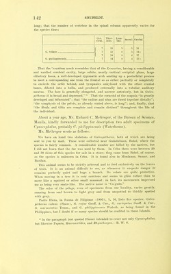
![The Philippine Journal of Science. D. General Biology, Ethnology and Vol. VI, No. 3, June, 1911. THE SKELETON IN THE FLYING LEMURS, GALEOPTERID/E. By R. W. Shufeldt. (Washington, D. C.) INTRODUCTION. Osteological mateiial for the present contribution has been furnished by Professor J. B. Steere of Ann Arbor, Michigan, and by Mr. Richard C. McGregor, of the Bureau of Science, Manila, P. I. What this material consists of, together with letters and other notes accompanying it, will be set forth further on in the present introduction. A number of comparative anatomists have touched upon the morphology of probably several of the species of the flying lemurs, but until the present time it appears that no fully illustrated and detailed account of the osteology of these remarkable animals has been published. Owen,^ in giving the characters of the skeleton of the Insectivora, briefly refers to the skull and some few of the limb bones of Galeopithecus, but in the case of the skull, unfortunately, he does not make it sufficiently clear as to whether the description does not likewise apply to Pteroptis. Thus he says: This [that is the skull] in Pteropus and Galeopithecus manifests the lissen- cephalous affinity by the squamosal being perforated by a venous canal behind the root of the zygoma, by the suspension of the malar, in the zygoma, by the distinct petrotympanic, by the vertical occiput, small cranial cavity, and blended orbital and temporal foss®. The orbit is partly defined behind by long and slender processes of the frontal, which is perforated by a superciliary foramen. The parietals usually coalesce at the sagittal suture, but rarely develop a crest. The paragraph continues in confused and inaccurate generalities to the end and closes with the statement that “in GaleopWiecm the coronoid is small.’’ Little more than this is added to his description of the skeleton, in fact only that In the Colugo (Galeopithecus) the ulna terminates in a point at the lower ^Anatomy of Vertebrates (1866), 2, 387, 388.](https://iiif.wellcomecollection.org/image/b22419020_0005.jp2/full/800%2C/0/default.jpg)