Anatomy, descriptive and surgical / by Henry Gray ; teh drawings by H.V. Carter ; with additional drawings in the second and later editions by Dr. Westmacott ; the dissections jointly by the author and Dr. Carter ; with an introd. on general anatomy and development, by T. Holmes.
- Henry Gray
- Date:
- [1877]
Licence: Public Domain Mark
Credit: Anatomy, descriptive and surgical / by Henry Gray ; teh drawings by H.V. Carter ; with additional drawings in the second and later editions by Dr. Westmacott ; the dissections jointly by the author and Dr. Carter ; with an introd. on general anatomy and development, by T. Holmes. Source: Wellcome Collection.
Provider: This material has been provided by the Francis A. Countway Library of Medicine, through the Medical Heritage Library. The original may be consulted at the Francis A. Countway Library of Medicine, Harvard Medical School.
52/974
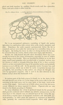
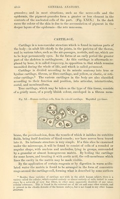
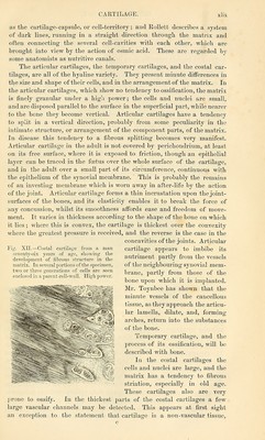
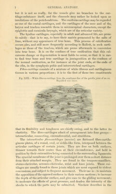
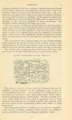
![but it is Bot so really, for the vessels give no branches to the car- tilage-substance itself, and the channels may rather be looked upon as involutions of the perichondriiim. The ensiform cartilage may be regarded a.s one of the costal cartilages, and the cartilages of the nose and of the_ larynx and trachea resemble them in microscopical characters, except the epiglottis and cornicula laryngis, which are of the reticular variety. The hyaline cartilages, especially in adult and advanced life, are prone to calcify—that is to say, to have their matrix permeated by the salts of lime, without any appearance of true bone. This process of calcification occurs also, and still more frequently according to EoUett, in such carti- lages as those of the trachea, which are prone afterwards to conversion into true bone. It is on the confines of true ossification that this cal- careous change or degeneration is most liable to occur, so that it is rare to find true bone and true cartilage in juxtaposition at the confines of the normal ossification, as for instance at the joint ends, at the ends of the ribs, in the symphysis pubis and intervertebral cartilages. Fihro-cartilage consists of a mixture of white fibrous and cartilaginous tissues in various proportions; it is to the first of these two constituents Fig. XIII.—White fibrous cartilage ti-om the semihinar disc of the patella joint of an o.x. Mao'nified loo times. that its flexibility and toughness are chiefly owing, and to the latter its elasticity. The fibro-cartilages admit of arrangement into four groups— interarticular, connecting, circumferential, and stratiform. The interarticular fibro-cartilages [menisci] are flattened fibro-cartila- ginous plates, of a round, oval, or sickle-like form, interposed between the articular cartilages of certain joints. They are free on both surfaces, thinner towards their centre than at their circumference, and held in position by their extremities being connected to the surrounding ligaments. The synovial membrane of the joint is prolonged over them a short distance from their attached margin. ^ They are found in the temporo-maxillary, sterno-clavicular, acromio-clavicular, wrist and knee joints. These car- tilages are usually found in those joints which are most exposed to violent concussions, and subject to frequent movement. Their use is—to maintain the apposition of the opposed surfaces in their various motions; to increase the depth of the articular surface, and give ease to the gliding movement; to moderate the effects of great pressure, and deaden the intensity of the shocks to which the parts may be submitted. Virchow describes in the](https://iiif.wellcomecollection.org/image/b21055087_0052.jp2/full/800%2C/0/default.jpg)