A treatise on the science and practice of midwifery / By W. S. Playfair.
- William Smoult Playfair
- Date:
- 1885
Licence: Public Domain Mark
Credit: A treatise on the science and practice of midwifery / By W. S. Playfair. Source: Wellcome Collection.
Provider: This material has been provided by the Augustus C. Long Health Sciences Library at Columbia University and Columbia University Libraries/Information Services, through the Medical Heritage Library. The original may be consulted at the the Augustus C. Long Health Sciences Library at Columbia University and Columbia University.
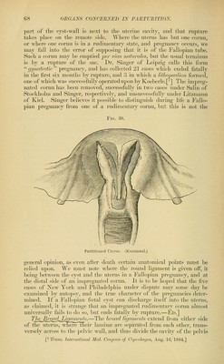
73/708 page 65
![They cease at the external orifice of the cervix, and they secrete the thick, tenacious, and alkaUne mucus which is generally found filling the cervical cavity. The transparent follicles, known as the '^ ovula Nabothii,'' w^hich are sometimes found in considerable numbers in the cavity of the cervix, consist of mucous follicles, the mouths of which have become obstructed and their canals distended by mucous secretion. The lower third of the cervical canal, as well as the exterior of the cervix, is covered with pavement epithelium; while on its upper portion is found a columnar and ciliated epithelium similar to that lining the uterine cavity. Bandl^ describes the cervical mucous membrance as extending much higher in the virgin than in women who have borne children, being traceable in the former nearly to the middle of the body of the uterus. During the first pregnancy he believes that the upper portion of the cervix is taken up into the body of the uterus, its mucous membrane never regaining the arrangement peculiar to that of the cervical canal. Vesseh^a[_JheUteriis.—The arteries of the uterus are derived from the intCTualillacanH'n'om ^^ ovarian. They enter the uterus between the folds of the broad ligaiuents^ and, penetrating its muscular coat, anas- tomose freely with each other and with the corresponding vessels of the opposite side. Their walls are thick and well developed, and they are remarkable for their very tortuous course, forming spiral curves, espe- cially in the upper part of the uterus. They end in minute capillaries which form the fine meshes surrounding the glands, and in the cervix give oif the loops entering the papillae. Beneath the uterine mucous membrane these capillaries form a plexus, terminating in veins without valves, which unite with each other to form the large veins traversing the substance of the uterus, known during pregnancy as the uterine sinuses, the walls of which are closely adherent to the uterine tissues. These veins, freely anastomosing with each other, pass outward to the folds of the broad ligaments, where they unite to form, with the ovarian and vaginal veins, a large and well-developed vejious network, known as the pampimfgrm plexus. ThelA^nj)Im^^ Uterus.—The lymphatics of the uterus are large'anHwelTTIevelopecr, ancTtliey have recently, and with much prob- ability, been supposed to play an important part in the production of certain puerperal diseases. A more minute knowledge than we at pres- ent possess of their course and distribution will probably throw much light on their influence in this respect. According to the researches of Leopold,^ who has studied their minute anatomy carefully, they originate in lymph-spaces between the fine bundles of connective tissue forming the l^sis of the mucous lining of the uterus. Here they are in intimate contact witli the utricular glands and the ultimate ramifications of the uterine l>lood-vessels. As they pass into tlie nmscular tissue tlicy be- come gradually narrowed into lymph-vessels and sjjaces, which have a very com])licated arrangement, and which eventually unite togetlier in the external muscular layer, especially on the sides of the uterus, to form large canals whi(;h probably have valves. Inmiediately under this ])eriton('al cov(-'ring these lymph-vessels foi-m a large; and t^hai'acteristic ' ArrJi.f. rjyn., V,. xiv. S. 2:57. = Il'i'l., 15(1. vi. Heft i.](https://iiif.wellcomecollection.org/image/b2121072x_0073.jp2/full/800%2C/0/default.jpg)