A treatise on the science and practice of midwifery / By W. S. Playfair.
- William Smoult Playfair
- Date:
- 1885
Licence: Public Domain Mark
Credit: A treatise on the science and practice of midwifery / By W. S. Playfair. Source: Wellcome Collection.
Provider: This material has been provided by the Augustus C. Long Health Sciences Library at Columbia University and Columbia University Libraries/Information Services, through the Medical Heritage Library. The original may be consulted at the the Augustus C. Long Health Sciences Library at Columbia University and Columbia University.
77/708 page 69
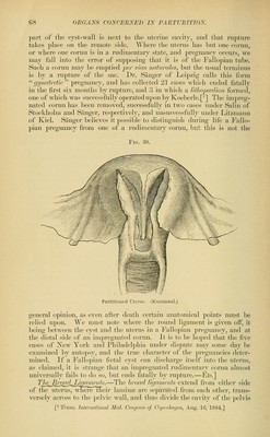
![into two parts; the anterior containing the bladder, the posterior the rectum. Their upper borders are divided into three subsidiary folds, the anterior of which contains the round ligament, the middle the Jlal- lopian tube, and thejjosterior the ovary. The arrangement has received the name of the ala vespertilioms, from its fancied resemblance to a bat's wing. Between the folds of the broad ligaments are found the uterine vessels and nerves, and a certain amount of loose cellular tissue con- FlG. Adult Parovarium, Ovary, and Fallopian Tube. (After Kobelt.) tinuous with the pelvic fasciae. Here is situated that peculiar structure called the organ of Rosenmiiller, or th.Q parovarium (Fig. 31), which is the remains of the Wolffian body, and corresponds to the epididymus in the male. This may best be seen in young subjects by holding up the broad ligaments and looking through them by transmitted light; but it exists at all ages. It consists of several tubes (eight or ten according to Farre, eighteen or twenty according to Bankes ^), which are tortuous in their course. They are arranged in a pyramidal form, the base of the pyra- mid being toward the Fallopian tube, its apex being lost on the surface of the ovary. They are formed of fibrous tissue, and lined with pave- ment epithelium. They have no excretory duct or communication with eitlier the uterus or^vary, and their function, if they have any, is un- known. MuHGidar Fibres behveen its Fojch.—A number of muscular fibres are also found in this situation, lying between the meshes of the connective tissue. They have been particularly studied by Rouget, who describes tliem as interlacing with each other, and forming an open network con- tinuous with the muscular tissues of the uterus (Fig. 32). They are (livisible into two layers, the cinterior of wliich is continuous with the nniscular fil)res of the; anterior surface of tiic uterus, and goes to form ]>art of the round ligament; the jMjsterior arises from th(! posterior wall of the uterus, and proceeds transversely outward to become attached to the sacTcviliac synchondrosis. A continuous mus(;ular enveh^pe is thus formed, which surrounds tlio whole of the uterus. Fallopian tubes, and ' Bankes, On iMn WolJJiau. BodieK.](https://iiif.wellcomecollection.org/image/b2121072x_0077.jp2/full/800%2C/0/default.jpg)