A treatise on the science and practice of midwifery / By W. S. Playfair.
- William Smoult Playfair
- Date:
- 1885
Licence: Public Domain Mark
Credit: A treatise on the science and practice of midwifery / By W. S. Playfair. Source: Wellcome Collection.
Provider: This material has been provided by the Augustus C. Long Health Sciences Library at Columbia University and Columbia University Libraries/Information Services, through the Medical Heritage Library. The original may be consulted at the the Augustus C. Long Health Sciences Library at Columbia University and Columbia University.
82/708 page 74
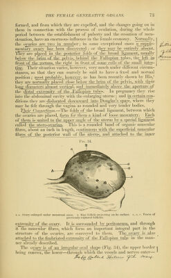
![being straiglit. 'J'lie anltxiur surfuiie, like ihat of the uterus, is less conxii2LJjjiULlllG_4X)stw^ The outer extremity is more rounded and bulbous than ihejniieii, \vliioh_ is somewhat poLuted^and eventually lost in jts4)njj2erJjgimient- By these peculiarities it is possible to distinguish the left from the right ovary after they have been removed from the body. The ovary varies much in size under different circumstances. On an average, in adult life, it measures from one to two inches in length, three-quarters of_an inch in_width, and about half an inch in thickness. It^jncreases greatly in size during each menstrual period— a fact Avhicli has been demonstrated in certain cases of ovarian hernia, in A\'liicli the j)rotruded ovary has been seen to swell as menstruation commenced; also during_j[)regnancy, when it is said to be double its usual size. Al^iMh£]^ange_j)fMife_jt_atiX2pl^^ and becomes rough and wrinkled on its surface. Before puberty, the surface of the ovary is smooth and polished and of a whitish color. After menstruation commences, its surface becomes scarred by the ruptur£jif_the r4rnafian follicles (Fig. 34, a ((), each of which leaves a little linear or striated cicatrix of a brownish color; and the older the patient the greater is the number of these cicatrices. Th^.;iX^ Stimc'turc—The structure of the ovary has been made the subject of many important observations. It has an_external covering of epithelium, originally continuous with the peritoneum, called by some the germ-epithelium, in consequence of the ovules being formed from it in early fcetal life. In the adult it is separated from the peri- toneum at the base of the organ by a circular white line, and it consists of colunuiar epithelium, differing only from the epithelium lining_jhe Fallopian tubes, with which it is sometimes continuous through the attached fimbria uniting the tube and the ovary, in_being^destitute^of cilia. Wmmediately beneath this covering is the dense coat known as the tunica alLuginea, on account of its w4iitish color. It consists of short connective-tissue fibres, arranged in laminae, among which are interspersed fusiform muscular fibres.^ At the point where the vessels and nerves enter the ovary this membrane is raised into a ridge, which is continiK)i3.a_witli the utei'o-ovnrian lioament, andjs called the hilmn. 'The tunjca albuginea is so intimately blended I'^'f'- '^'5. with the stroma ofrl^ ovary as to be insep- arable on dissection ; it\loes not, however, e_x- ist as a distinct lamina, but is merely the ex- ternal part-of the proper structure of the ovary, in which more dense connectijiejtissue is de- veloped than elsewhere. The Stroma.—On making a long-itudinal sec- tion of^the ovary (Fig. 35), it will be seen to be composed of two parts, the more internal of which is of a reddish color from the number of vessels that ramify in it, and is called the mecJuUw'ii or vascular zone; while the e^cter- iiaj^'of a whitisir~tinf7reccives the name of the (^r/Zm/^orjx^^ The former consists of loose connective tissue niterspersed^witlTelastic, and a considerable number of Longitudinal Seotion of Adult Ovary. (After I'arre.)](https://iiif.wellcomecollection.org/image/b2121072x_0082.jp2/full/800%2C/0/default.jpg)