A treatise on the science and practice of midwifery / By W. S. Playfair.
- William Smoult Playfair
- Date:
- 1885
Licence: Public Domain Mark
Credit: A treatise on the science and practice of midwifery / By W. S. Playfair. Source: Wellcome Collection.
Provider: This material has been provided by the Augustus C. Long Health Sciences Library at Columbia University and Columbia University Libraries/Information Services, through the Medical Heritage Library. The original may be consulted at the the Augustus C. Long Health Sciences Library at Columbia University and Columbia University.
83/708 page 75
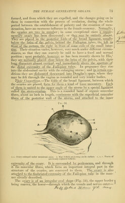
![M ^ ' / muscular fibres. According to Rouget^ and His,^ the muscular structure forms the greater part of the ovarian stroma. The latter describes it as consisting essentially of interwoven muscular fibres, which he terms the fusiform tissue, and which he believes to be continuous with the muscular layers of the ovarian vessels. The former believes that the muscular fasciculi accompany the vessels in the form of sheaths, as in erectile tissues. Both attribute to the muscular tissues an important influence in the expulsion of the ovules and in the rupture of the Graafian fol- FiG. 36. ' / '■ >-. .-> --::, </// '^>rt '-B-:- -■- Section through the Cortical Part of the Ovary, e. Surface epithelium, s s. Ovarian stroma. 1 1. Large-sized Graafian follicles. 2 2. Middle-sized; and 3 3. Small-sized Graafian follicles, o. Ovule within Graafian follicle, v v. Blood-vessels in the stroma. </. Cells of the membrana granulosa. (After Turner.) licles. Waldeyer and other writers, however, do not consider it to be so extensively developed as Rouget and His believe. The cortical sub-1 stance is the more important, as that in which the Graafian follicles and I ovules are formed. It consists of interlaced fibres of connective tissue, containing a large number of nuclei. The muscular fibres of the medul- lary substance do not seem to penetrate into it in the human female. In it are fi)und the Graafian follicles, Avhich exist in enormous numbers from the earliest periods of life, and in all stages of development (Fig. 36.) TIie^m(if/m -Fo/^'c/eg.^r-According to the researches of Pfliiger, Waldeyer, and other German writers, the Graafian follicles are formed in early foetal life by (cylindrical inflections of the epithelial, covering of the ovary, which di]) into the substance of the gland. These tubular filaments anastomose witli each otlier, and in tliem are formed tlie ovules, which are originally the epithelial cells lining the tubes. Por- tions l)Cconi(; shut off from the rest of the fihiinents and fi>rm the Graafian fidlicles. The ovules, on this view, are highly-developed epithelial cells, originally derived from the surface of the ovary, and ' Jon.rn/il. de. PIi.i/kIo/., i. p. T?)?. ^ Schiiltze'.s Arch.f. jMikrn/^c.np. Anal., 1865.](https://iiif.wellcomecollection.org/image/b2121072x_0083.jp2/full/800%2C/0/default.jpg)