Manual for the physiological laboratory / by Vincent Dormer Harris and D'Arcy Power.
- Harris, Vincent Dormer.
- Date:
- 1888
Licence: Public Domain Mark
Credit: Manual for the physiological laboratory / by Vincent Dormer Harris and D'Arcy Power. Source: Wellcome Collection.
Provider: This material has been provided by the Augustus C. Long Health Sciences Library at Columbia University and Columbia University Libraries/Information Services, through the Medical Heritage Library. The original may be consulted at the the Augustus C. Long Health Sciences Library at Columbia University and Columbia University.
11/280 (page 3)
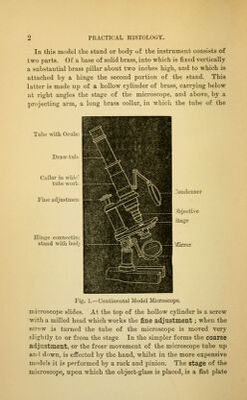
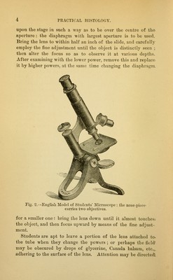
![of brass blackened upon its upper surface, or of glass cemented on to a (lark background, and perforated by a central aperture. Through the aj)crturc light is reflected from a mirror ; the amount of light admitted to the lens is regulated by a small cylinder which fits into the central aperture; this, when withdrawn from below, will receive the diaphragms ; these are perforated by apertures of different sizes ; the smallest is a mere pinhole, and is for use with the strongest magnifying ])0wer. One of the diaphragms should be inserted into the cylinder, and re- placed in the centre of the stage. When in position it is exactly flush with the upper surface. In some microscopes this form of diaphragm is replaced by a blackened disc of metal perforated near its circumference by holes of various sizes, and revolving round its centre. At the back of the stage is a pair of brass clips for holding the slides in position when the microscope is tilted. In some instruments the movements of the stage in different directions are effected by screws; but in this the stage is fixed, and the finger and thumb suffice to move the slide upon it. Beneath the stage is a movable mirror with two faces, one concave, the other plane ; the concave mirror is the more com- monly used, as it condenses the light upon the object and thus affords a better illumination. The tube of the microscope consists of a hollow brass cylinder from five to eight and a half inches in length. It contains a second or draw-tube, which can, if necessary, be drawn out, in order to increase the magni- fying power. The ujjper extremity of the tube receives the ocular or eye-piece : the oculars vary in magnifying power. It is better not to change the oculars, but always to w'ork with one of moderate power. Into the lower end of the tube the objectives, powers, oi lenses should be screwed. These also vary in magnifying power. In using the microscope, i)ut on a low power first; then adjust the mirror in such a way as to get a full illumination of the field, so that the eye applied to the ocular sees a circle of light of equal intensity in all its parts. Never use the direct rays of the sun. Next put the object to be examined 1—2](https://iiif.wellcomecollection.org/image/b21219618_0011.jp2/full/800%2C/0/default.jpg)