Glioma retinae : with report of five cases / by Christian R. Holmes.
- Christian Rasmus Holmes
- Date:
- [1902]
Licence: In copyright
Credit: Glioma retinae : with report of five cases / by Christian R. Holmes. Source: Wellcome Collection.
Provider: This material has been provided by UCL Library Services. The original may be consulted at UCL (University College London)
20/36 page 18
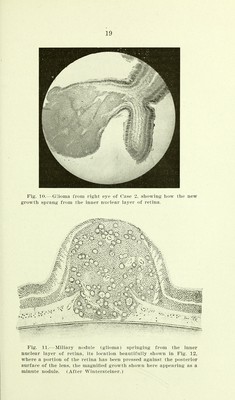
![current growth springing from the optic nerve: The structure is that of small round cells in definite connec- tion with a fihrous intercellular substance. The blood vessels are poorly formed and pigment granules are found in many parts. The tumor is a small round- celled sarcoma. Case 2.—Baby R. E., aged 5 months, was brought to me Dee. 29, 1897. The child was well developed and nourished. Three weeks ago the parents noticed that the pupil of the right eye was dilated, and had a silvery appearance which made the eye look dull. Dr. W. F. Shepherd was consulted, and promptly referred the case for consultation. The globe was congested; T.=-l-I; cornea clear; anterior chamber ob- literated; lens and iris in contact with cornea; pupillary space filled with a mass of a reddish gray color, the surface being unusually \ ascular, the disease having advanced to the second stage. The appearance of the growth when examined under a strong light, gives a peculiar reflex not unlike a fire opal. Ophthalmoscopic examination of the left eye under mydriasis was negative. The child was admitted to my hospital; the same day enucleation with extensive resection of the optic nerve performed, and the specimen given to Dr. H. J. Whitacre for microscopic examination. The following is his report: The specimen received was a complete right eye, which was placed in a 4 per cent, solution of formol for four days, and the hardening completed in 95 per cent, alcohol. A sagittal section of the hardened specimen presented the appearance shown by the photograph (Fig. 6), and demon- strates a globular tumor occupying mainly the temporal half of the vitreous chamber. Posteriorly, the nasal half shows a pedicle attached to the point of entrance of the optic nerve. Anteriorl}^, the tumor presses against the posterior surface of the lens, and on each side of the detached retina can be seen a thin membrane extending from the tumor to the choroid. The tumor, when half hardened, was very soft, and must have been almost gelatinous in its fresh state. Sections of the tumor were made with dilliculty because of the multiple points of calcareous degeneration which dulled the knife and tore the sections. In general arrangement the tumor is made up of bands and circles of cells arranged regularly about the central opening (Fig. 7). The appearance is that of a vascular tumor with the cells concentrically arranged around the central vessel. ]iy higher magnification the lumen of this central vessel looks like the acinus of a gland surrounded by high columnar cells (Fig. 8) ; the other cells of the area have similar char- acteristics because of their origin by proliferation from these](https://iiif.wellcomecollection.org/image/b21637611_0022.jp2/full/800%2C/0/default.jpg)