Practical anatomy : a manual of dissections / by Christopher Heath.
- Christopher Heath
- Date:
- 1902
Licence: In copyright
Credit: Practical anatomy : a manual of dissections / by Christopher Heath. Source: Wellcome Collection.
Provider: This material has been provided by The University of Leeds Library. The original may be consulted at The University of Leeds Library.
37/782 (page 13)
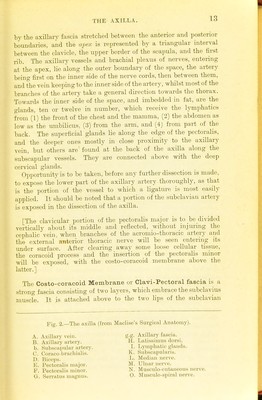
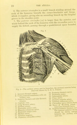
![by the axillary fascia stretched between the anterior and posterior boundaiies, and the apex is represented by a triangular interval between the clavicle, the upper border of the scapula, and the first rib. The axillary vessels and brachial plexus of nerves, entering at the apex, lie along the outer boundary of the space, the artery being first on the inner side of the nerve cords, then between them, and the vein keeping to the inner side of the artery, whilst most of the branches of the artery take a general dii-ection towards the thorax. Towards the inner side of the space, and imbedded in fat, are the glands, ten or twelve in number, which receive the lymphatics from (1) the fi'ont of the chest and the mamma, (2) the abdomen as low as the umbilicus, (3) from the arm, and (4) from part of the back. The superficial glands lie along the edge of the pectoralis, and the deeper ones mostly in close proximity to the axillary vein, but others are found at the back of the axilla along the subscapular vessels. They are connected above with the deep cervical glands. Oppoi-tunitj-is to be taken, before any further dissection is made, to expose the lower part of the axillary artery thoroughly, as that is the portion of the vessel to which a ligature is most easily applied. It should be noted that a portion of the subclavian artery is exposed in the dissection of the axilla. [The clavicular portion of the pectoralis major is to be divided vertically about its middle and reflected, without injmmg the cephalic vein, when branches of the acromio-thoracic artery and the external anterior thoracic nerve will be seen entering its under sm-face. After clearing away some loose cellular tissue, the coracoid process and the insei-tion of the pectoralis minor will be exposed, with the costo-coracoid membrane above the latter.] The Costo-coracoid Membrane or Clavi-Pectoral fascia is a strong fascia consisting of two layers, which embrace the subclavius muscle. It is attached above to the two lips of the subclavian Fig. 2.—The axilla (from MacliHo's Surgical Anatomy). A. Axillary vein. B. Axillary artery. 1). Subscapular artery. C Coraco-bracliialis. D. Biceps. K. Pectoralis nuijor. P. Pectoralis minor, ti. Serratus niagnuB. g.g. Axillary fascia. H. Latissimus dorsi. L Lymphatic glands. K. Subscapularis. Ii. Median ner\-e. M. Ulnar nerve. N. Museulo-c^utaneous nerve. O. Musculo-si)iral nerve.](https://iiif.wellcomecollection.org/image/b21508562_0037.jp2/full/800%2C/0/default.jpg)