The principles and practice of obstetric medicine and surgery : in reference to the process of parturition / by Franics H. Ramsbotham.
- Francis Henry Ramsbotham
- Date:
- 1865
Licence: Public Domain Mark
Credit: The principles and practice of obstetric medicine and surgery : in reference to the process of parturition / by Franics H. Ramsbotham. Source: Wellcome Collection.
Provider: This material has been provided by the Harvey Cushing/John Hay Whitney Medical Library at Yale University, through the Medical Heritage Library. The original may be consulted at the Harvey Cushing/John Hay Whitney Medical Library at Yale University.
50/778 page 48
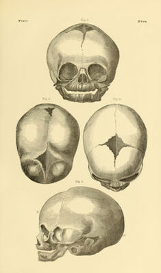
![48 OFTHEFCETALHEAD. bregmatic (/, c), ?>h inches. These measurements, though different from those generally stated by the various obstetrical writers, are now generally accepted in this country. They are the results obtained by Prof. Meigs, after an accu- rate examination of one hundred and fifty heads ; and, as such, deserve the con- fidence of every American practitioner. We have ourselves examined over seventy-five heads, and our observations are such as to convince us of the cor- rectness of Prof. Meigs' statement. It is true that our average for the bi-parie- tal was about 4| inches, and of the occipito-mental 5 J inches; but the difference in the number examined might account for this discrepancy. The fact, how- ever, is positive, that the measurements of the foetal head, as laid down by Euro- pean writers, will not answer for this country. The attachment of the head to the spinal column is an important item in obstetrics ; for, as the result of this arrangement, we have one of the acts of the mechanism of labour, viz , flexion, by means of which in most cases the vertex becomes the presenting point of the foetal head. The articulation of the head with the atlas is nearer the occipital extremity of the occipito-mental diameter. If the head, resting upon the spine, represent a lever, and the propelling force be exerted through the vertebral column, the point of resistance being at the extremities of the occipito-mental diameter, then, in parturition, the occipital extremity of the head should descend first. This is generally so, and doubtless the above is the explanation.] Anatomical Peculiarities of the Foetal Skull.—The general anatomical character, as well as the form and size of the skull, deserve our attention. It may be seen that the bones are not dovetailed into each other as in the adult, but are separated to some extent by intervening lines and spaces of membranous formation. The lines are termed sutures, from the Latin word suo, to sew ; the spaces, fontanelles, after the French, because it used to be supposed that a moisture distilled from the brain through these unossi- fied apertures. The fontanelle has also been called bregma, from /3ps'xw, to moisten—the name having originated in the same idea. The bones in the child's skull requiring our consideration obstetrically are but few:—the two parietal bones of a square shape, which give the principal protection to the brain laterally (plate 5, fig. 4, a, b); the frontal bone ante- riorly (plate 5, fig. 1)—or rather the frontal bones, because, in the foetus there are two ;—and the occipital posteriorly (plate G). The parietal bones are separated from the frontal, or connected with them, by a suture called coronal (plate 5, fig. 3, and plate 5, figs. 1 and 2), which runs from near the external angle of one eye to the same point on the opposite side of the head, bounding the forehead superiorly. It is called coronal, because the ancients used to wear their coronse or garlands on that part of the head upon festive occasions. The parietal bones are separated from the occipital by a suture, termed lamdoidal, from its resemblance to the Greek letter A, (plates 6 and 5, fig. 2). The two parietal bones are separated from each other by the sagittal suture (plate 5, fig. 4) which runs longitudinally along the centre of the upper part of the head, so called, because it was fancifully supposed to be situated between the lamdoidal and coronal sutures, as an arrow is placed in a strung bow. The two frontal bones are separated by the frontal suture (plate 5, fig. 1), which runs directly upwards from the root of the nose. The remaining sutures of the head are out of the way of our obstetrical observation, and a description of them would therefore be useless. The two fontanelles are placed, one at each extremity of the sagittal suture; and they are named, according to their situation, anterior (plate 5,](https://iiif.wellcomecollection.org/image/b21007123_0050.jp2/full/800%2C/0/default.jpg)