Licence: Public Domain Mark
Credit: Exsection of the hip-joint / by C.T. Poore. Source: Wellcome Collection.
Provider: This material has been provided by the Augustus C. Long Health Sciences Library at Columbia University and Columbia University Libraries/Information Services, through the Medical Heritage Library. The original may be consulted at the the Augustus C. Long Health Sciences Library at Columbia University and Columbia University.
14/36 page 8
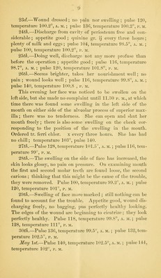
![three inches below the trochanter; tliis hius continued to dis- chari^e profusely ever since, ller mother can assign no cause for the trouble. On examination, there is found a sinus as above, from which there is a profuse discharge of healthy-look- ing })us. Both limbs are plump, and no difference in their size can be detected ; there is no pain along the shaft of the femur on pressure. The right thigh is flexed somewhat on the abdomen. Crepitus can be detected by crowding the head of the bone inward ; no exposed bone can be found. Pa- tient is a blonde, and seems to be in good condition; appetite fair. Extension applied. April 21, 1875,—Patient was to-day etherized, and, as- sisted by i)r. Watts, I made the usual incision over the trochanter, and exposed the bone. In attempting to throw the head of the bone out of the acetabulum the femur was fractured at the junction of the diaphysis with the e[)iphysis, just above the knee-joint. Tlie head of the bone was re- moved from the acetabulum after section, just above tro- chanter minor. On examination of the cut end of the bone, it was found to be soft, discolored, the periosteum being easily separated, and on crowding the end of the bone up in order to see the extent of the disease, the same condition was found. It remained either to amputate at the hip-joint, or to remove the whole shaft of the femur : as the patient was apparently in good condition, the latter was decided upon. This was easily done, the whole shaft being drawn bv traction through the original incision. There was no haemorrhage, nor was the periosteum attached at any point. The fracture was found to have taken place obliquely from within outward and from behind, forward, leaving a small portion of the ex- ternal condyle attached to the epi])hysis, but dead. The ace- tabulum was perforated; the upper portion of the wound was brought together with suture, leaving a sufficient opening for tlie discharge; no involucrum had formed. Tlie limb was placed between sand pillows, and extension (four pounds) ap- plied. 8 p. M., patient comfortable, does not complain of any pain ; pulse 128 and good ; temperature 10(>.5°. 22<:7.—Comfortable ; temperature 10<>.5°, pulse 13(3, a. m. ; temperature 100.5°, pulse 142, r. m.](https://iiif.wellcomecollection.org/image/b21210640_0014.jp2/full/800%2C/0/default.jpg)




 No text description is available for this image
 No text description is available for this image
 No text description is available for this image