The American text-book of operative dentistry / In contributions of eminent authorities. Ed. by Edward C. Kirk.
- Edward Cameron Kirk
- Date:
- 1900
Licence: Public Domain Mark
Credit: The American text-book of operative dentistry / In contributions of eminent authorities. Ed. by Edward C. Kirk. Source: Wellcome Collection.
Provider: This material has been provided by the Augustus C. Long Health Sciences Library at Columbia University and Columbia University Libraries/Information Services, through the Medical Heritage Library. The original may be consulted at the the Augustus C. Long Health Sciences Library at Columbia University and Columbia University.
102/864 (page 100)
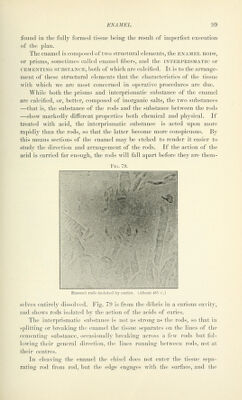
![force applied at an acute angle with the direction of the rods fractures the tissue in the lines of least resistance. If the edge be keenly shar]), it will enter the tissue slightly, and then th(! bevel acts as a wedge in addition to the force applied to the shaft of the instrument; but if the edge be dull, it will rest across the ends of many rods, will not engage with the surface, and the force applied will break and crumble the tissue but will not cleave it. The enamel rods, or prisms, are long, slender prismatic rods or fibers, five- or six-sided, pointed at both ends and alternately expanded and constricted throughout their length. They are from 3.4 to 4.5 microns^ in diameter, some of them apparently reaching the entire distance from the surface of the dentin to the surface of the enamel ; but as the diameter of the rods is the same at their outer and inner ends, and as the crown surface is much greater than the surface of den- tin covered by enamel, there are many rods which do not extend through the entire thickness. These short rods end in tapering points between the converging rods which extend the entire distance. To express this in terms of development: as the formation of enamel begins at the surface of the dentin, the increasing area of crown sur- face requires more ameloblasts, and as new ameloblasts take their place in the layer the formation of new enamel rods begins between the rods which were previously forming. These short rods are most numerous over the marginal ridges and at the points of the cusps, and will be considered more fully in connection with those positions. In ground sections cut at right angles to the direction of the rods ^ the tissue has the appearance of a mosaic floor, the outline of the rods being more distinct if they have been marked out by treating the section slightly with a(nd (Fig. 80). In longitudinal sections (Fig. 81) the sides of the rods are not smooth and even like the sides of a lead pencil, but are alternately expanded and constricted. They are well illustrated by taking balls of soft clay and sticking them together one above another to form a rod, then putting a number of rods together so that by mutual pressure they take hexagonal forms. This illustrates also the manner of growth of the tissue in formation. The expansions and constrictions can be seen in rods that have been scraped from a cleaved surface of enamel, but better by isolating rods by the slight action of dilute acid (Fig. 82). In the construction of the tissue the rods are so arranged that the ex- ' A micron is tlie unit of microscopic measurement, and is equal to one one-thousandtli of a millimeter. ^ In describino; the direction of enamel rods they are always considered as extending from the dentin to the surface, and the angle is formed at the surface of the dentin with the locating plane, either horizontal or axial.](https://iiif.wellcomecollection.org/image/b21216629_0102.jp2/full/800%2C/0/default.jpg)