The American text-book of operative dentistry / In contributions of eminent authorities. Ed. by Edward C. Kirk.
- Edward Cameron Kirk
- Date:
- 1900
Licence: Public Domain Mark
Credit: The American text-book of operative dentistry / In contributions of eminent authorities. Ed. by Edward C. Kirk. Source: Wellcome Collection.
Provider: This material has been provided by the Augustus C. Long Health Sciences Library at Columbia University and Columbia University Libraries/Information Services, through the Medical Heritage Library. The original may be consulted at the the Augustus C. Long Health Sciences Library at Columbia University and Columbia University.
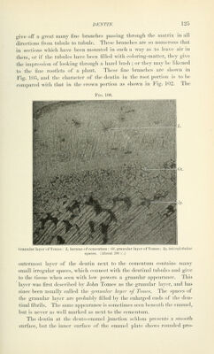
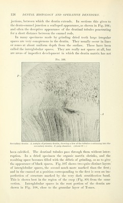
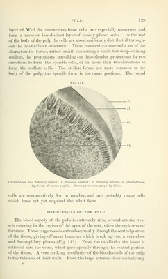
129/864 (page 127)
![The formation of dentin is not complete at the time of eruption of the tooth, but continues for an indefinite period, thickening the layer of dentin at the expense of the pulp. When the typical amount of den- tin has been formed the growth ceases, and does not begin again unless excited by some irritation to the pulp or the pulp of some other tooth of the same side, whi(;h leads to the formation of secondary dentin. Secondary dentin is never as perfect in structure as primary dentin; the tubules are smaller, fewer, and much more irregular. Often in ground sections several periods of formation can be determined by dif- ferences of structure, each deposit becoming successively more and more imperfect in structure. This is shown in Fig. 109. Pulp. The dental pulp is the soft tissue occupying the central cavity of the dentin. It is made up of embryonal connective tissue and contains a large number of bloodvessels and nerves. Like all connective tissues, the intercellular substance is large in amount and the cells are widely scattered in tliis soft, jelly-like tissue, which contains but few fibers. We recognize four kinds of cells in the pulp : the odontoblasts, form- ing the outer surface of the pulp next to the dentin; and round, spindle- shaped, and stellate connective-tissue cells. AREAXGEMENT OF CELLS. The odontoblasts are tall columnar cells, sometimes club-shaped, and in older tissues, which have ceased to be functional, sometimes becoming almost spherical. They form a continuous layer over the entire surface of the pulp, being everywhere in contact with the dentin. The layer has been called the membrana eboris, or the membrane of the ivory. The nuclei of the odontoblasts are large and oval, containing a large amount of chromatin, and are very different from the nuclei of ordinary connective-tissue cells. Three kinds of processes have been described in connection with the odontoblasts : 1. The dentinal fibril processes, or fibers of Tomes. These are long, slender ])rotoplasmic processes projecting from the dentin end of the cell into a dentinal tubule, and running through the tubule to the outer surface of the dentin. Usually there is but one fibril extendino; from each odontoblast, but sometimes two can be seen, extending into two tubules. These fibrils can be demonstrated in decalcified sections or Ijy removing the pulp from a recently extracted tooth by cracking the tooth and carefully lifting the pulp out of the pulp chamber, and then either teasing or sectioning. Fig. 110 shows the fibrils projecting from](https://iiif.wellcomecollection.org/image/b21216629_0129.jp2/full/800%2C/0/default.jpg)