The American text-book of operative dentistry / In contributions of eminent authorities. Ed. by Edward C. Kirk.
- Edward Cameron Kirk
- Date:
- 1900
Licence: Public Domain Mark
Credit: The American text-book of operative dentistry / In contributions of eminent authorities. Ed. by Edward C. Kirk. Source: Wellcome Collection.
Provider: This material has been provided by the Augustus C. Long Health Sciences Library at Columbia University and Columbia University Libraries/Information Services, through the Medical Heritage Library. The original may be consulted at the the Augustus C. Long Health Sciences Library at Columbia University and Columbia University.
86/864 (page 84)
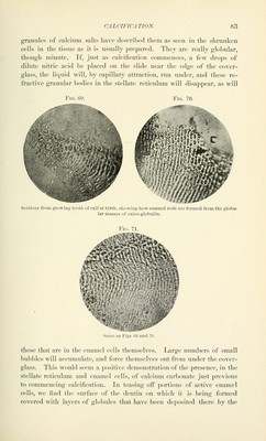
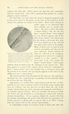
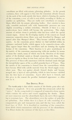
![eiiamol cells (Fij;. 68). These, given (nit from tli(> cell continually, form the cnaniel rods. One rod is separated tVoni anotlici' hy a |n-oto- j)lasnuc cenieut substance. Dr. Graf Spee says that whcii the tissue is j)roj)erly ])repared—and he lays great stress on this point—at the time of the formation of the enamel, the globules are always to be found. Their entire absence at „ „„ earlier stap-es is an indication that Fig. 72. ^ these globules are an enamel sub- stance. He gives to them the name '* enamel drops, and says he saw these enamel drops, when enamel is to be formed, appear only in the half of the enamel cells wdiich rests [J on tiic dentin; afterward they were to be seen farther up in the cell, but not quite to the region of its nucleus. Many of them were so small as to be scarcely measurable, and they are al- ways spherical. Great numbers of Section of (leveinpiM t .1, of calf at birth, ^\^^,^ ^^^ collected at the periphery, showinj; tirst-foriiiiii;; layer i)f enamel. The , l i j 7 globules of caico-giobuiin are seen arranged and appear here either to be completely in lines where rods are to be formed: 1 ^^y^^^^ ^.^ to fuse together. The enamel cells containing caleo-spherites; ■:!, ^ b globules arranged to form rods; 3, first- loWCr part of the cell COntainS the forming layer of enamel: 4, dentin. 1 ,, ^ ^ )5 1 • 1 larger enamel drops, which merge without sharp boundaries into the substance of the enamel rods. This then appears as a part of the enamel cell, in which the originally iso- lated enamel drops have run together into a continuous mass, and the growth of the enamel rod, once begun, appears to take place by the addition of new enamel drops. The minute globular forms described by Dr. Sj^ee are calco-s])herites ; the larger ones, his enamel drops, are globules of calco-globulin which are to form the rods (Fig. 72). Appearances of calcified fibers ])roj('cting beyond the line of calci- fication are seen when studying sections of forming young enamel, and these are evidences that fine processes of fibers from the cells of the stratum intermedium pass down through and among the ameloblasts to the forming enamel beneath. These are probably the processes which i\rr. Tomes saw and descrilied as connecting the enamel cells with the stratum intermedium. If one separates slightly the enamel cells from the stratum intermedium the j^arted cells will have the appearance of broken processes or fibers, and we may be able to see fibers crossing from the enamel cells to the stratum intermedium. A longitudinal section of a human tooth at birth, just after the](https://iiif.wellcomecollection.org/image/b21216629_0086.jp2/full/800%2C/0/default.jpg)