The American text-book of operative dentistry / In contributions of eminent authorities. Ed. by Edward C. Kirk.
- Edward Cameron Kirk
- Date:
- 1900
Licence: Public Domain Mark
Credit: The American text-book of operative dentistry / In contributions of eminent authorities. Ed. by Edward C. Kirk. Source: Wellcome Collection.
Provider: This material has been provided by the Augustus C. Long Health Sciences Library at Columbia University and Columbia University Libraries/Information Services, through the Medical Heritage Library. The original may be consulted at the the Augustus C. Long Health Sciences Library at Columbia University and Columbia University.
92/864 (page 90)
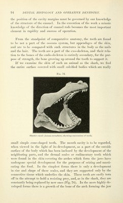
![In a recent ]);i|){'r on tlie Histology of the Pulp, by Erwin Hoelil, he states that the cells of the pulp show in the diU'erent life periods cha- racteristic differences in form and number. Three kinds are found, Avhieh arise from one another by metamorphosis in the following way : (1) Round cells with large nucleus and scanty ])rot(ii)lasm. (2) Irregu- larly shaj)ed cells with many freely anastouiosing ])roccsses. (.'i) Spindle- sha])c(l cells with the same character as the ibregoing. The (changes of the cell form begin at the })eriphery and ])roceed towai'd the centre of the ])ulp. The outermost ])eri])heral layer of the branched cells contains the elementary or primary odontoblasts. Ccntralward from these is a cell layer which, with reference to the function of its elements, is called the conjugation cell layer. The secondary odontoblasts arise by conjugation of the primary odontoblasts with the conjugation cells, and they form the dentin. The conjugation processes probably cease only with the com])letion of growth in the tooth. Of the peripheral processes of the primary odontoblasts the larger one represents what will be the future dentin fibril. The increase of cells seems to be dependent upon the development of the capillaries, inasmuch as more cells are found where the distribution of capillaries is most dense, i. e. on the periphery of the pulp. Tlie gradual decrease of the number of branch cells in the centre of the pulp during the course of development is because only trunk vessels are found here. In the place of these destroyed cells we find a delicate cellular network which is probably derived from the numerous anastomoses of the cell processes. Next to or just within the odontoblastic layer is seen a bright zone variable in width ; this is the so-called Weil's layer. Between this and the fibrous or central portion of the pulp is an intermediate layer which forms a contrast with the delicate fibrous elements of Weil's layer, and in this way Weil's layer is made visible. The ground substance of the pulp by a certain method of treatment shows a dense interlacing of fibrillse wdiieh are arranged parallel to one another and seem to run in the direction of the axis of the tooth. The Gum. Gum tissue is the same as that of the general mucous membrane of the mouth. It is more dense because it is bound down to the bone by numerous fibers of its own, and it is also united with the periosteal tissue which spreads into it in every direction. Numerous large single and compound papillffi are seen. The blood supply is abundant, but nerve tissue is not often found. The histological appearances which look like young enamel organs are the glands of Serres. Near develop- ing teeth epithelial clusters are frequently seen, the remains of the dis- appearing necks of the enamel organs. The cells of the stratum Mai-](https://iiif.wellcomecollection.org/image/b21216629_0092.jp2/full/800%2C/0/default.jpg)