The American text-book of operative dentistry / In contributions of eminent authorities. Ed. by Edward C. Kirk.
- Edward Cameron Kirk
- Date:
- 1900
Licence: Public Domain Mark
Credit: The American text-book of operative dentistry / In contributions of eminent authorities. Ed. by Edward C. Kirk. Source: Wellcome Collection.
Provider: This material has been provided by the Augustus C. Long Health Sciences Library at Columbia University and Columbia University Libraries/Information Services, through the Medical Heritage Library. The original may be consulted at the the Augustus C. Long Health Sciences Library at Columbia University and Columbia University.
98/864 (page 96)
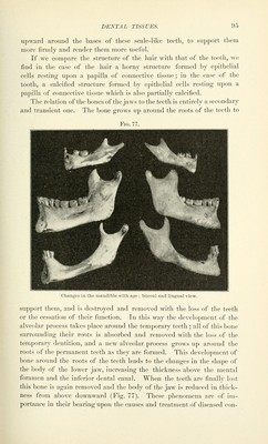
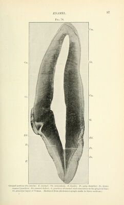
![ditions of the teeth, particuhirly those which involve the supporting tissues. Dental Tissues.—The human teeth arc made up of four tissues (Fig. 78): 1. The enamel covers the exposed portion of the tooth, or crown, and gives the detail of crown form. Its function is to protect the tooth against the wear of friction. 2. The dentin forms the mass of the tooth and determines its class form, the number of cusps and the number of roots being indicated by the dentin form. 3. Cementinn covers the dentin beyond the border of the enamel, overlapping it slightly at the gingival line and forming the surface of the root. Its function is to furnish the attachment of the fibers of the ])eridental membrane, which fastens the tooth to the bone. 4. The jjulp or soft tissue filling the central cavity in the dentin is the remains of the formative organ which has given rise to the dentin. Its functions are the formation of dentin and a sensory function. In describing the structure of the teeth and the arrangement of the structural elements of the tissues directions are described Avith reference to three planes : The mesio-disto-axial plane, a plane passing through the centre of the crown from mesial to distal and parallel with the long axis of the tooth. The bucco-linguo-axial plane, a plane passing through the centre of the crown from buccal to lingual and parallel with the loiig axis of the tooth. The horizontal plane, at right angles to the axial planes. The Supporting Tissues.—The human teeth are supported on the maxillary bones, their alveolar processes growing up around the roots of the teeth, so that the roots fit into the holes in the bone. The calcified structures of the tooth and the bone are not, however, united, but the roots are surrounded by a fibrous membrane, the jjer-iclental membrane, or fte^'ic^inentum, which fastens the tooth to the bone. Enamel. The enamel differs from all other calcified tissues in the nature of the structural elements of which this tissue is made up, in the degree of calcification, and in origin, being the only calcified tissue derived from the epiblast. The enamel is formed from an epithelial organ derived from the epithelium of the mouth cavity and indirectly from the epiblastic germ layer, while all other calcified tissues are products of the mesoblast. In the case of bone and dentin the formative tissue is persistent. It](https://iiif.wellcomecollection.org/image/b21216629_0098.jp2/full/800%2C/0/default.jpg)