Relations of diseases of the eye to general diseases : forming a supplementary volume to every manual and text-book of practical medicine and ophthalmology / by Max Knies ; edited by Henry D. Noyes.
- Date:
- 1895
Licence: Public Domain Mark
Credit: Relations of diseases of the eye to general diseases : forming a supplementary volume to every manual and text-book of practical medicine and ophthalmology / by Max Knies ; edited by Henry D. Noyes. Source: Wellcome Collection.
Provider: This material has been provided by the Royal College of Physicians of Edinburgh. The original may be consulted at the Royal College of Physicians of Edinburgh.
20/492 (page 2)
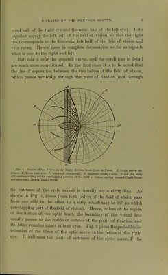
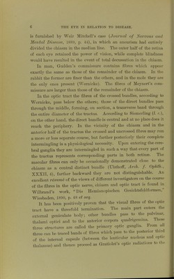
![A. Anatomical Course of the Nerves of the Eye. We are to consider the optic nerve, the nerves of the ocular mus- cles, the trigeminus, facial, and sympathetic nerves. Of these by far the most important is 1. The Optic Nerve. This collects the centripetal and centrifugal fibres from the nerve- fibre layer of the retina, undergoes partial decussation in the chiasm L A Fig 1 -Field of Vision of Both Eyes (Schematic). X. Left; R, right half of the field of vision, divided by the vertical line ab which passes through the point of fixation F. The ver- tical strip is the overlapping portion of the field of vision. with the nerve of the opposite side, and sends fibres through the optic tracts to both cerebral hemispheres. The fibres from the inner (nasal) half of the retina (corresponding to the outer [temporal] half of the field of vision) form the larger part of the nerve and undergo decussation. The fibres from the outer half of the retina, and which answer to the nasal side of the field, pass without decussation to the cerebral hemisphere of the same side. The optic tract thus contains all the fibres from the halves of the retinse on the same side, the right tract containing the fibres of the right retinal halves (the tern-](https://iiif.wellcomecollection.org/image/b21969188_0020.jp2/full/800%2C/0/default.jpg)