A manual of instruction in the principles of prompt aid to the injured : including a chapter on hygiene and the drill regulations for the hospital corps, U.S.A. : designed for military and civil use / by Alvah H. Doty.
- Alvah Hunt Doty
- Date:
- 1898, ©1894
Licence: Public Domain Mark
Credit: A manual of instruction in the principles of prompt aid to the injured : including a chapter on hygiene and the drill regulations for the hospital corps, U.S.A. : designed for military and civil use / by Alvah H. Doty. Source: Wellcome Collection.
Provider: This material has been provided by the Francis A. Countway Library of Medicine, through the Medical Heritage Library. The original may be consulted at the Francis A. Countway Library of Medicine, Harvard Medical School.
43/336 (page 19)
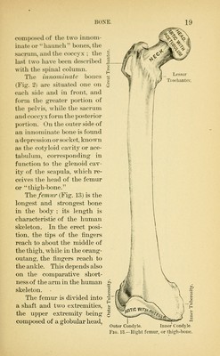
![HONK (M)ni]Miscil of the two iiuioin- iiiatc or liatiiicli Woiies, the Siicrum, aiul the coceyx ; the last two have been described with the spinal cohunn. The uino)ni)ia(e bones (Fig. 2) are situated one on each side and in front, and form the greater portion of the pelvis, while the sacrum and coccyx form the posterior porticm. On the outer side of an innominate bone is found a depression or socket, known as the cotyloid cavity or ace- tabulum, corresponding in function to the glenoid cav- ity of the scapula, which re- ceives the head of the femur or ''thigh-bone. The femur (Fig. 13) is the longest and strongest bone in the body ; its length is characteristic of the human skeleton. In the erect posi- tion, the tips of the fingers reach to about the middle of the thigh, while in the orang- outang, the fingei^ reach to the ankle. This depends also on the comparative short- ness of the arm in the human skeleton. The femur is divided into a shaft and two extremities, the upper extremity being composed of a globular head, Lesser Trochanter. Outer Condyle. Inner Condyle. Fig. 13.—Right femur, or thigh-bone.](https://iiif.wellcomecollection.org/image/b21049543_0043.jp2/full/800%2C/0/default.jpg)