Lectures on the development of the gravid uterus / by William O. Priestley.
- Priestley, William Overend, 1829-1900.
- Date:
- 1860
Licence: Public Domain Mark
Credit: Lectures on the development of the gravid uterus / by William O. Priestley. Source: Wellcome Collection.
Provider: This material has been provided by The Royal College of Surgeons of England. The original may be consulted at The Royal College of Surgeons of England.
60/114 page 56
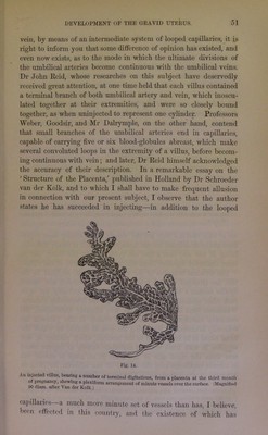
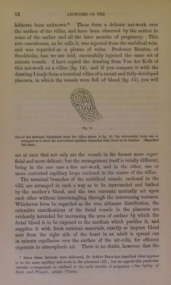
![conveyed into tliese cells or spaces between the ramifications of the umbilical vessels ; and that, after having acted upon the child’s blood through the coats of the latter, it was subsequently returned into the nterine veins or sinuses. The later researches of Owen, and the report of Messrs Stanley and Mayo on the preparations in the Hunterian Museum, all tended to confirm the correctness of these observ'ation.s. Weber and Dr John Eeid, while they concur in the general views of the Hunters, concerning the double cir- culatioii in the placenta, hold that the utero-placental vessels are ]>rolonged beyond the layer of decidua lying on its surface, into the substance of the organ. According to the former, the delicate inner coat only of the materaal vessels penetrates the after-birth, and forms there a large vascular net-work, ramifying in the intervals of the placental tufts, and forming large sinuses into which the villi project, carrying the walls of the .sinuses before them. Dr Reid de.scribes the maternal placenta to consist of a large .sac formed by the inner coat of the vascular system of the mother, which is intersected in many directions by the placental tufts, the latter projecting into it like fringe.s, and pushing its thin wall before them in the form of sheaths, which closely envelop both the tnink and each individual branch composing these tufts. Blood is brought by the maternal arteries to this .sac, and returned from it without extravasation by the utero-placental veins. Dr Reid even saw foetal tufts penetrating soine of the sinuses situated in the uterine walls, and beyond the exact limits of the outer surface of the placenta. Mr Dalrymple subseqiiently denied the existence of any maternal cells in the placenta, and stated that simple spongy interspaces were present among the villi into which the mother’s blood was projected. The researches of Eschricht and M. Bonami, detailed by M. Cazeau, led them to conclude that the maternal vessels pass through the decidua, and form in the substance of the placenta a net-work of exceedingly delicate meshes, which ramify and embrace everywhere the tufts of the umbilical vessels enclosed in the chorial villi Schroeder van der Kolk, who had, during a cholera epidemic, excellent opportunities in Holland for making investigations on this subject, expresses his conviction that the decidua observed on the uterme surface of the placenta sends down into the substance of the organ dissepiments, which penetrate even to its foetal surface, and circumscribe .spaces, wherein the foetal villi are suspended, and into which the matenial blood is poiired from the uterine arterie.s, to pa.ss thence into the uterine](https://iiif.wellcomecollection.org/image/b22334452_0062.jp2/full/800%2C/0/default.jpg)