Volume 1
A manual of operative surgery / by Sir Frederick Treves.
- Sir Frederick Treves, 1st Baronet
- Date:
- 1903
Licence: Attribution-NonCommercial 4.0 International (CC BY-NC 4.0)
Credit: A manual of operative surgery / by Sir Frederick Treves. Source: Wellcome Collection.
Provider: This material has been provided by The University of Leeds Library. The original may be consulted at The University of Leeds Library.
694/808 (page 674)
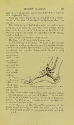
![and the long one below it. The ankle-joint lies about on the level of a point half an inch above the tip of the inner malleolus. The position of the tendons about the ankle-joint must be borne in mind, as also the situation of the tibial and peroneal arteries. The lower epiphysis of the tibia includes the articular sur- face and the internal malleolus. Ossification coimiiences in it during the second year, and the epiphysis joins the shaft be- tween the eighteenth and nineteenth years. The lower epi- physis of the fibula includes the articular surface and outer malleolus. Ossification commences in the second year, and is completed about the twenty-first year. Both epiphyseal lines are horizontal, and are brought in contact with that pouch of synovial membrane Avhich extends upwards between the tibia and fibula. Operation.—The various methods in vogue for performing this operation are, for the most part, modifications of the original procedure of Moreau. Indeed, no very conspicuous deviations from the initial operation have been proposed or carried out. Of the modern forms of Moreau's operation, that by Langenbeck would appear to be one of the best. It may be carried out as follows, if the subperiosteal method be attempted:— The patient lies upon the back, with the foot and leg su])- ported lipon a firm sand pillow. Two vertical lateral mcisions are made. 1. The Outer Incision.—The foot being turned over upon its inner side, a vertical incision some three inches in length is made along the anterior part of the fibula to a point a little below the tip of the malleolus. Thence it is made to curve around the malleolus, and ascend for about one inch along its posterior border (Fig. 201, b). 2. The Removal of the Fibida.—The fibula is exposed, and its periosteum divided in the long axis of the bone. The membrane is then separated from the bone by the rugine in an anterior and a posterior direction. The ligaments attached to the malleolus are separated as encountered. The external lateral ligament is divided vertic^illy, so that its anterior segment will go with the](https://iiif.wellcomecollection.org/image/b21511342_0001_0696.jp2/full/800%2C/0/default.jpg)