The journal of anatomy and physiology : normal and pathological. Vol. XV. [Pt. 4] / conducted by G.M. Humphry [and others].
- Date:
- 1881
Licence: Public Domain Mark
Credit: The journal of anatomy and physiology : normal and pathological. Vol. XV. [Pt. 4] / conducted by G.M. Humphry [and others]. Source: Wellcome Collection.
Provider: This material has been provided by The Royal College of Surgeons of England. The original may be consulted at The Royal College of Surgeons of England.
14/148 page 458
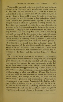
![Dalton,1 Beigel,2 Slavjansky,3 and others, have minutely described these degenerate follicles. On July 15, 1880, Dr Bantock removed a large multilocular cyst of the right ovary from a robust young married woman, aged 28, whom we will briefly term “ Case 2.” She had been married, nearly two years, and not having menstruated for several months, was believed to be pregnant. The left ovary was clearly in a state of incipient cystic degeneration, and. was therefore removed. It measured 2| inches in its long diameter, and weighed 360 grains. Examined fresh, the surface was but little puckered; on section it appeared full of thin-walled cysts containing highly albuminous fluid. These cysts were simply Graafian follicles bearing perfect ova. Between these follicles the stroma was abundant, pale, and succulent. On microscopic examination no morbid changes could be detected in the tunica albuginea. The stroma consisted of the normal spindle-shaped cells, not closely packed, but freely dis- tributed as in a healthy child’s or young girl’s ovary. This we noted to counteract any hasty conclusions that cirrhotic changes in the stroma may prevent the follicles from bursting, and allow them to slowly develop into large cysts. There were plenty of cysts but no cirrhosis. In fact, the loose stroma was over- abundant, and bore large numbers of degenerate follicles (by this term we imply such as have never taken a share in preg- nancy nor menstruation). Some were reduced to a mere cloudy sinuous band, no longer forming a complete chain; the tissue partly included by this band was looser than elsewhere, yet not undergoing any kind of degeneration. Most of these follicles formed fusiform bodies composed of radiating cloudy tubes, or rather bands, bearing traces of degenerate nuclei. In the centre were broken-down masses of pigment. The tubes were, ex- ternally, very sharply bordered by the surrounding stroma, which, however, sent in filiform processes of elongated and nucleated cells (see fig. 3). b ‘1 Report on the Corpus Luteum,” Trans. Amcr. Gynecological Soc. vol. ii. 1877. 3 “ Zur Naturgeschichte des Corpus Luteum,” Archiv. f. Gyncvkologic, vol. xiii, 1878. * “Zur norma]en und Pathologisclien Histologie des Craafschen Blaschens des Mensehen,” Virchow’s Archiv. vol. li. 1870.](https://iiif.wellcomecollection.org/image/b22455875_0016.jp2/full/800%2C/0/default.jpg)


 No text description is available for this image
 No text description is available for this image
 No text description is available for this image