The blood-corpuscle considered in its different phases of development in the animal series / by T. Wharton Jones.
- Thomas Wharton Jones
- Date:
- 1846
Licence: Public Domain Mark
Credit: The blood-corpuscle considered in its different phases of development in the animal series / by T. Wharton Jones. Source: Wellcome Collection.
Provider: This material has been provided by The Royal College of Surgeons of England. The original may be consulted at The Royal College of Surgeons of England.
12/50 (page 72)
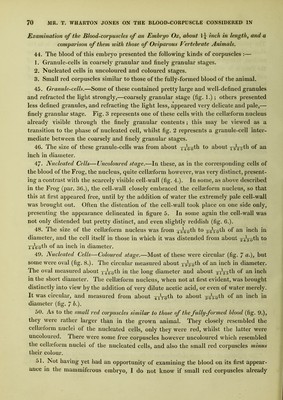
![Frog (par. 27.). In this state they look as if they had burst, but such is not the case in general. 59. By the addition of water, the collapsed granule-cells become distended and their appearance is thus distinctly brought out. Fig. 1 a. represents the human gra- nule-cell in its coarsely granular stage thus distended ; the contained granules, it will be observed, are minute, and on attentive observation they are seen to be in active molecular motion. Fig. 2 b. represents the finely granular stage of granule-cell of human blood, distended by water. 60. The granule-cell of human blood is sometimes seen to present, though not very strikingly, a clear spot indicating, as I think, the place of the nucleus, similar to that above represented in the case of the granule-cell of the Skate. 61. When distended by the action of water the human granule blood-cell is about 2-4-ooth of an inch in diameter. 62. Fig. 3 a. represents the granule-cell, coarsely granular stage, and fig. 4 the granule-cell in its finely granular stage of the blood of the Horse. Figs. 5 a. and 6 represent the coarsely and finely granular stages of the granule-cell of the blood of the Elephant. It will be observed that the diameter of these cells is somewhat greater than that of the granule-cell of human blood, but the principal difference is the size of the contained granules in the coarsely granular stage. Whilst in the granule-cell, coarsely granular stage, of the Horse the granules may be estimated in round numbers at about T-g- \-Ath of an inch in diameter, those in the same cell of the Ele- pliant are about ]0QQth, and those in the human granule blood-cell about 25 oOQth. 63. If, after the granule blood-cell has been distended by the action of water, very dilute acetic acid be added, the contained granules are dissolved, and a cellseform nucleus brought into view. Fig. 1 b. represents a human granule-cell so acted on ; fig. 3 b. a granule blood-cell of the Horse similarly acted on; and fig. 5 b. a granule blood-cell of the Elephant in the process of being acted on by acetic acid. This last drawing was made before the granules were all dissolved, but when the cellseform nucleus was already distinctly in view. In the case of the cells here represented, as indeed in the case of many others, I watched the progress of the action of the acetic acid on the granules and the coming into view of the cellseform nucleus. 64. If, before the addition of the acid, the granule-cell has not been distended by the action of water, and if the acid has not been much diluted, instead of one cellse- form nucleus of the size represented, and which in the human granule-cell may be about 0th of an inch in diameter, but larger in the Elephant and smaller in the Horse, an appearance of several smaller ones variously aggregated may be brought out. This, however, in opposition to what is generally believed, and in opposition to what I myself once believed, I can affirm most positively is merely an appearance artificially produced by the corrugating action of the acid on the walls of the single celleeform nucleus, altogether in the manner above shown to be the case with the cellaeforra nucleus in the corresponding cells of the blood of the Frog.](https://iiif.wellcomecollection.org/image/b22290564_0014.jp2/full/800%2C/0/default.jpg)