Volume 1
Cyclopaedia of obstetrics and gynecology / edited by Egbert H. Grandin.
- Grandin, Egbert H. (Egbert Henry), 1855-
- Date:
- 1889
Licence: Public Domain Mark
Credit: Cyclopaedia of obstetrics and gynecology / edited by Egbert H. Grandin. Source: Wellcome Collection.
Provider: This material has been provided by the Royal College of Physicians of Edinburgh. The original may be consulted at the Royal College of Physicians of Edinburgh.
45/544 page 29
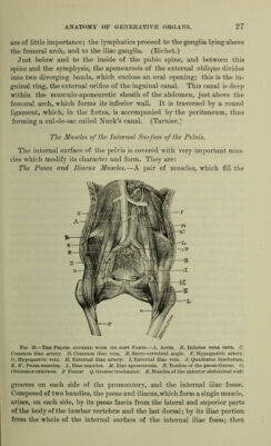
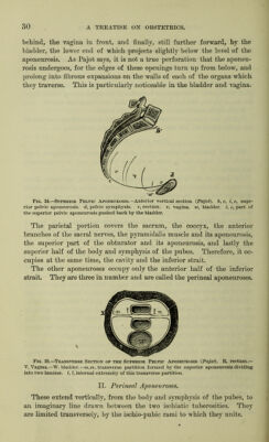
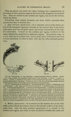
![it is carried forward, and, for a distance of seven to eight inches, crosses the muscles to an orifice which forms the anus. The lateral parts of the sacrum, externally to the greater sciatic notch, and the anterior sacral canals, are occupied by the pyramidalis muscle. It stretches over the osseous spaces which separate the anterior sacral canals, across the greater sciatic notch, to the internal surface of the great trochanter. The anterior branches of the sacral nerves, which form the sacral plexus, the hypogastric artery and vein, cross this muscle. A true aponeurosis covers the pyramidalis muscle. Finally, the aponeurotic and peritoneal lamina cover those portions of the cavity which are not covered by either muscles or viscera. Floor of the Pelvis, We shall borrow the greater part of our anatomical description from Dubois and Pajot, for ip. their work this intricate part is most clearly explained. The floor of the pelvis, inferior wall of the pelvis, perineal floor, perineum, is formed by a muscular aponeurotic plane, ])ierced by three openings, the anus, the vulva and the orifice of the urethra, which open on its surface. It forms an elastic couch, which plays an important part in delivery. Two very distinct parts can be distinguished: an aponeurosis, formed of layers which unite and make distinct spaces, and muscles, which fill these spaces and are accompanied by important vessels and nerves. Su'perior Pelvic Aponeurosis. {Figs, 24 and 25.) Proceeding from within outwards, the first aponeurosis is the superior pelvic aponeurosis. Attached behind to the anterior surface of the sacrum and coccyx, inside to the sacral canals, and in front on the internal sur- face of the body of the pubes, near the symphysis, it extends along the entire wall of the pelvis, excepting the superior half of the anterior semi- circumference. On a level with the edge of the abdominal strait, it blends in front with the aponeurosis of the abdominal walls; laterally with the iliac muscle; behind with the lumbo-iliac aponeurosis. Below it covers the entire floor of the pelvis like a kind of inferior diaphragm. On a level with the superior border of the great sciatic notch, it divides into two laminae which, twisting at almost a right angle, form a trans- verse partition that divides this aponeurotic cavity into two parts, the anterior, which is the larger, and the posterior. These two laminae, joined by their superior edge, turn aside at their inferior part, thus form- ing a triangle, base downward. Their internal portion meets the rectum and vagina, with the walls of which they blend, their external portion reaching the soft parts, wliicli occupy the ischiatic canal. The perineal portion is crossed by the rectum](https://iiif.wellcomecollection.org/image/b21704867_0001_0045.jp2/full/800%2C/0/default.jpg)