Licence: Public Domain Mark
Credit: Ovum / by Allen Thomson. Source: Wellcome Collection.
Provider: This material has been provided by The University of Glasgow Library. The original may be consulted at The University of Glasgow Library.
133/150 (page 129)
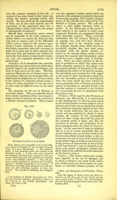
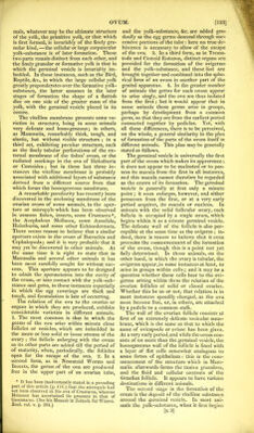
![near the anterior extremity of the cell. A number of ova were found in the ovary con- taining the distinct germinal vesicle with macula. He also observed the segmentation of these ova in the usual manner, and the conversion of the segmented mass into a ciliated embryo, within which the new polype is subsequently developed.-]' Should these observations prove correct and be applicable to the other instances of similar winter ova among the Bryozoa, they may tend to remove some of the difficulties which exist in regard to the various repro- ductive bodies occurring in these animals ; but farther researches seem still necessary to point out in these and in other polypine ani- mals more fully and minutely the relation be- tween the three kinds of reproductive bodies, viz., true ova, separated gemmules, and at- tached buds. Acalephce.—It is remarkable that notwith- standing the very close relation in which these animals stand to the Anthozoid Polypes, the form of their ova is not the same. The Dis- cophora (Medusae) are of distinct sexes: the Ctenophora (Beroes) are hermaphrodite ; the Siphonophora (Diphyidae) are various, or bear, in the manner of compound animal stocks, a variety of zoids, sometimes of one sex alone, at other times of different sexes on the same stem. The structure of the ova in Medusae is extremely simple. They are originally formed from minute cytoblasts which soon acquire a single nucleus or macula, and are enclosed in a delicate external membrane. These consti- Fig. 95*. Development of the ova of Acalepha. These figures give magnified views of the diffe- rent stages of formation of the ova taken from the ovary of a large Rhizostoma. a. The primitive germ. 6. The germinal vesicle now present in the primitive ovum. c. d. The same more advanced and enlarged, the macula has appeared in the ger- minal vesicle, and a few yolk granules are deposited in the clear vitelline substance, e. The yolk gra- nules greatly increased in quantity and becoming opaque, a vitelline membrane is now formed, f The same somewhat more advanced, the yolk gra- nules are now collecting together to form cor- puscles. The macula is assuming the elongated form. t Proceedings of British Association for 1855. See also Professor Allman’s interesting Report on the Polyzoa to the British Association. See Trans, for 1850, p. 320. tute the germinal vesicles, round which the granular yolk-substance is gradually deposited in increasing quantity. The complete segmen- tation of the yolk has been observed by Von Siebold in Cyanea aurita.* The yolk-sub- stance is often highly coloured, violet or yellow. In the former part of this article I have referred to the manner in which some compound Hydroida are propagated through their medusoid progeny. These medusoid individuals, like the ordinary Medusae, are of separate sex; and they must therefore be looked upon as the complete stage of the polypine animals from which they have proceeded, whether they have their }'Oung developed while the parent remains at- tached to the nursing polype stock, or have assumed the separate and independent mode of life in a more complete state of develop- ment. There are many varieties in the de- gree of perfection to which they attain even while remaining attached to the polype; but the general principle of formation is the same throughout the whole of the hydroid animals, the remarkable and constant fact with regard to the mode of their reproduction being this, that the immediate product of development from the ovum which has been formed by sexual generation from a Medusa or medusoid animal is invariably an attached Polype, and that the medusa or medusoid is the product of a non-sexual process of gemmation from this polype stem. Protozoa.—With regard to the Protozoa, or Infusoria and Bhizopoda, it is unnecessary to add anything here to what has been stated in the several articles on these subjects and in a former part of this one, excepting the remark, that continued researches appear to show that as the sexual distinction has not been de- tected, and may probably be absent in these animals, the nucleus of the monocellular forms of these beings may hold the place of the germinal vesicle in them, and that the processes of division and production of in- ternal gemmules takes the place of true ovu- lation. At the same time it must be admitted that it is by no means improbable that the sexual relations may yet be discovered in the lowest monocellular animal bodies, as has re- cently been the case in some of the simpler and monocellular Algas, and that as our knowledge of the process of reproduction in these beings is still very limited, it may be destined to un- dergo even greater progressive changes than those which it has suffered from the researches of the last few years.-]' Porifera, — The bodies which have usually been regarded as the ova of Sponges, and to which a reference was made in the earlier part of this article, are of two kinds, viz. gem- mules or detached ciliated portions of the * Beitr. zur Naturgesch. der Wirbellos. Thiere, 1839. t See the papers of Focke, Cohn, and Stein re- ferred to in the first part of this article, and the more recent work of Stein, “ Die Infusionsthiere auf ihre Entwickelungsgeschichte untersucht.” 4to. Leipzig, 1854.](https://iiif.wellcomecollection.org/image/b24918751_0135.jp2/full/800%2C/0/default.jpg)