History of infusoria, including the desmidiaceae and diatomaceae, British and foreign / by Andrew Pritchard.
- Andrew Pritchard
- Date:
- 1861
Licence: Public Domain Mark
Credit: History of infusoria, including the desmidiaceae and diatomaceae, British and foreign / by Andrew Pritchard. Source: Wellcome Collection.
Provider: This material has been provided by Royal College of Physicians, London. The original may be consulted at Royal College of Physicians, London.
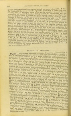
984/1074 (page 964)
![D.RSCU1PTI0N 01 THE ENOHAVINGS. PLATE XL. (EoTATOttiA). Figure 1. Hydatina senta, female, lateral view: a, dorsum and oral cavity, extending to an apex at 6; c, mastax with maxilla3; d, canal between mastax and stomacli; /, cloaca! orilice; g, vesicle; h, ovary; i, coils of respiratory tube; k, cerebral ganglion; I, ciliated tactile fossa; m, longitudinal muscles. 2. Enteroplea Hydatina, the male of Hydatina senta. 3. Ova in an immatiu-e state, as found in the unimpregnated ovary of Hydatina senta: a, germinal spot; b, germinal vesicle; c, membrane of ovum occupied with granular yelk-matter. 4. The lining membrane of stomach of Hydatina senta, everted, showing cilia. 5. Vibratile tag, supported on its pedicle, attached to the respiratory canal. 6. The male sexual organs (of Enteroplea Hydatina) detached, and highly magnified: a, penis; b, gland smTounding its bag; c, vesicles with granules; d, fold of integument siu-rounding penis when retracted. 7. Detached spermatozoa. 8. Stephanops muticus, seen from beneath. 9. Same, side view. 10. Another view from beneath, or the ventral surface. 11. Brachionus Dorcas, female, newly born. 12. Same, male, newly born (Gosse). 13. B. Miilleri (male): a, head mass; b, eye; c, muscles; d, posterior mass; e, sperm- sac ; /, urinary concretion; g, foot. 14. B. Pala, male, neMy born. 15. Same, male egg, nearly mature. 16. B. Bakeri. 17. Saeeulus viridis, male, newly born. 18. Same, female, with male ova attached. 19. Brachionus angularis, male. 20. B. urceolaris, mastax and dental apparatus, ventral aspect: a, mastax; b, malleus; c, manubrium; d, articulation; e, imcus ; /, incus; g, ramus; h, fulcrum; i, muscle connecting the imcus with the ramus; j, muscle for extending the malleus; I, muscle for thi'owing in the manubrium; k, muscle for bending the malleus; in, buccal funnel; n, salivary glands; 0, alula. [These letters have the same signification where met with in the following figures after Gosse:] 21-23. B. urceolaris: 21. Jaws viewed nearly from above ; 22. Dental apparatus, lateral aspect; 23. Buccal funnel, salivary glands, mastax, and dental appa- ratus, dorsal aspect. 24. Diglena forcipata, jaws closed, ventral aspect. 25. Floscularia ornata, jaws, dorsal aspect. 26. The same, frontal aspect. 27. Stephanoceros Eicli- hornii, jaws, dorsal aspect. 28. Same, uncus, oblique aspect.](https://iiif.wellcomecollection.org/image/b22652164_0986.jp2/full/800%2C/0/default.jpg)