Twelve lectures on the structure of the central nervous system for physicians and students / Trans by Willis Hall Vittum. Edited by C. Eugene Riggs.
- Ludwig Edinger
- Date:
- 1891
Licence: Public Domain Mark
Credit: Twelve lectures on the structure of the central nervous system for physicians and students / Trans by Willis Hall Vittum. Edited by C. Eugene Riggs. Source: Wellcome Collection.
Provider: This material has been provided by UCL Library Services. The original may be consulted at UCL (University College London)
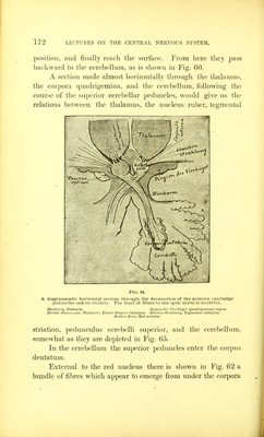
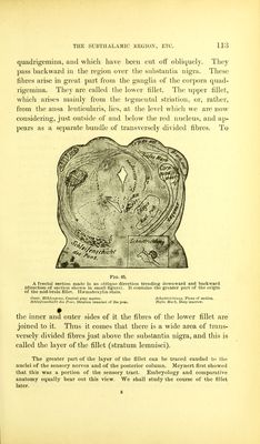
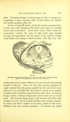
127/284 page 111
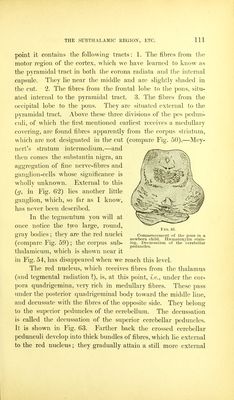
![point it contains tlie following tracts: 1. The fibres from the motor region of the cortex, which we have learned to know as the pyramidal tract in both the corona radiata and the internal capsule. They lie near the middle and are slightly shaded in the cut. 2. The fibres from the frontal lobe to tlie pons, situ- ated internal to the pyramidal tract. 3. The fibres from the occipital lobe to the pons. They are situated external to the pyramidal tract. Above these three divisions of tlie pes pedun- culi, of which the first mentioned earliest receives a medullary covering, are foinid fibres apparently from the corpus striatum, which are not designated in the cut (compare Fig. 50),—Mey- nert's stratum intermedium,—and then comes the substantia nigra, an aggregation of fine nerve-fibres and ganglion-cells whose significance is wholly unknown. External to this {g, in Fig. 62) lies another little ganglion, which, so far as I know, has never been described. In the tegmentum you will at once notice the two large, round, gray bodies; they are the red nuclei Commencement of the pons in a „. rrv\ 1 1 newborn child. Hsematoxylin stain- (compare rig. 59): the corpus sub- Decussation of the cerebellar ^ ^ o / ' 1 peduncles. thalamicum, which is shown near it in Fig. 54, has disappeared when we reach this level. The red nucleus, which receives fibres from the thalamus (and tegmental radiation]), is, at this point, under the cor- pora quadrigemina, very rich in medullary fibres. These pass under the posterior quadrigeminal body toward the middle line, and decussate with the fibres of the opposite side. They belong to the superior peduncles of the cerebellum. The decussation is called the decussation of the superior cerebellar peduncles. It is shown in Fig. 63. Farther back the crossed cerebellar pedunculi develop into thick bundles of fibres, which lie external to the red nucleus; they gradually attain a still more external](https://iiif.wellcomecollection.org/image/b21271264_0127.jp2/full/800%2C/0/default.jpg)