Elementary text-book of zoology. Special part: Mollusca to man / by C. Claus ; translated and edited by Adam Sedgwick ; with the assistance of F.G. Heathcote.
- Carl Friedrich Wilhelm Claus
- Date:
- 1897
Licence: Public Domain Mark
Credit: Elementary text-book of zoology. Special part: Mollusca to man / by C. Claus ; translated and edited by Adam Sedgwick ; with the assistance of F.G. Heathcote. Source: Wellcome Collection.
180/356 page 178
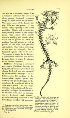
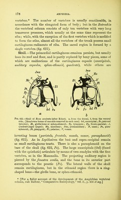
![vertebrso.* The number of vertebrse is usually considerable, in accordance with the elongated form of body; but in the Batrachia the vertebral column consists of only ten vertebrse with very long transverse processes, which usually at the same time represent the ribs; while, with the exception of the first vertebra which is modified to form the atlas, almost all the vertebrse of the trunk possess small cartilaginous rudiments of ribs. The sacral region is formed by a single vertebra (fig. 621). Skull.—The primordial cartilaginous cranium persists, but usually loses its roof and floor, and is partly replaced by bony pieces, some of which are ossifications of the cartilaginous capsule (exoccipitals, auditory capsules, sphen-ethmoid, quadrate), while others are a Fig. 622.—Skull of Rana esculenta (after Ecker), a, from the dorsal, b, from the ventral side; [Membrane bones of one side removed in each case]. Ocl, exoccipital; Pe, petrosal (prootic); Ft, girdle-bone or sphen-ethmoid; Ty, tympanic ; Fp, fronto-parietal; J, quadrato-jugal (jugal); Mx, maxillary; Jmx, prasmaxillary; N, nasal; Ps, para- sphenoid ; Pt, pterygoid; FI, palatine; V, vomer. investing bones (parietals, frontals, nasals, vomer, parasphenoid) (fig. 622). As in Lepidosiren the basi- and supra-occipital remain as small cartilaginous tracts. There is also a parasphenoid on the base of the skull (fig. 622, Ps). The large exoccipitals (Ocl) (fused with the opisthotic) articulate by means of two condyles with the first vertebra, as in the Mammalia. The projecting auditory region is pierced by the fenestra ovalis, and the bone in its anterior part corresponds to the prootic (Pe). The lateral walls of the skull remain cartilaginous, but in the ethmoid region there is a ring- shaped bone—the girdle bone, or sphen-ethmoid. * [For a fuller account of the development of the Amphibian vertebral column, vide Balfour, “ Comparative Embryology,” vol. ii., p. 456 et seq.J](https://iiif.wellcomecollection.org/image/b2813378x_0180.jp2/full/800%2C/0/default.jpg)