Elementary text-book of zoology. Special part: Mollusca to man / by C. Claus ; translated and edited by Adam Sedgwick ; with the assistance of F.G. Heathcote.
- Carl Friedrich Wilhelm Claus
- Date:
- 1897
Licence: Public Domain Mark
Credit: Elementary text-book of zoology. Special part: Mollusca to man / by C. Claus ; translated and edited by Adam Sedgwick ; with the assistance of F.G. Heathcote. Source: Wellcome Collection.
195/356 page 193
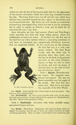
![Reproduction takes place in the spring. Copulation is confined to an external approximation of the two sexes, and almost always takes place in the water. The male, which sometimes has a wart-like elevation on the thumb (Ranch) or gland on the arm (Pelobates) embraces the female from the back, usually behind the front limbs, and pours out the seminal fluid over the spawn as it issues in strings or in clumps. The individual eggs are surrounded by a viscous layer of albumen which swells up in the water. The upper half of the ovum is of a darker colour than the lower. The process of segmentation begins in the upper part, and the con- strictions which lead to the formation of the segmentation spheres proceed more rapidly in this region than at the lower pole. With the end of segmentation a cavity—the segmentation cavity—appears in the mass of cells; it is placed nearer to the upper pole than to the specifically heavier lower pole. The germ [blastoderm], with medullary plate and folds, arises on the upper half; it quickly, even before the closure of the medullary canal to form the medullary tube, grows round the yolk. After development of the branchial arches and before the mouth is formed, the embryos which have a short tail leave their egg membranes as tadpoles at a stage of development which varies with the species. They then attach themselves by means of two suckers to the gelatinous remains of the spawn (similar suckers are present on the throat of the Triton-larvae, where however they are stalked). Most larvae leave the egg membranes with more or less developed rudiments of three pairs of branched external gills (vol. i., fig. 111). The body gradually increases in length and the fin-like tail developes. Later the mouth is formed and the larva begins to feed. Soon the external branchial appendages disappear, while the skin grows over the gill slits like an operculum in such a manner that only one gill aperture is left, through which the water flows out of the branchial chambers on either side. During these processes fresh lancet-shaped gill-plates are developed in double rows along each branchial arch. The mouth is armed with a horny beak, which is used in gnawing vegetable an 1 also animal substances. The intestine has become very long and much coiled, and the lungs have grown out of the pharynx in the form of long sacs. As development proceeds the hinder extremities first make their appearance on the body of the Tadpole close to the attachment of the strongly-developed swimming tail. As the pulmonary respi- ration increases, the branchial apparatus becomes more and more reduced, and the animal undergoes an ecdysis, with which is con- VOL. II. 13](https://iiif.wellcomecollection.org/image/b2813378x_0195.jp2/full/800%2C/0/default.jpg)