Licence: Public Domain Mark
Credit: A system of dental surgery / by Sir John Tomes. Source: Wellcome Collection.
22/808 page 6
![Behind the alveoli for the first temj^orary molars we have a large open socket, which, in the upper maxilla, has but a very imperfect posterior wall. Projecting inwards from the free edge of the outer and inner alveolar walls, wo may observe small spicula, the rudiments of septa which are destined to divide the cavity into two distinct sockets, and thus separate the pulps of the second temporary and first 2:)ermanent molar teeth, both of which at present occupy one large alveolus. The division usiially takes place a little earlier in the lower than the upper jaw. The groove whicli marks the jjassage of the nerve and artery in the floor of the socket of the first temporary molar, can be traced through the alveoli of the two posterior teeth, back to the inferior dental foramen, which is situated midway between the angle of the jaw and the edge of the inner wall of the alveolus of the first permanent molar, a little below the floor of the jjosterior part of the last alveolus. At this period the articiUar jDrocess of tlie lower jaw is scarcely raised above the level of the alveolar edge, while the angle is projected downwards a little below the general level of the inferior margin of the jaw. The coronoid \wo- cess rises at an angle of forty-five degrees from the alveolar edge, its ascent commencing at the anterior boundary of the socket of the first permanent molar. In the upper jaM' the zygomatic process proceeds outwards from the anterior margin of the large open socket of the second temporary molar. It is necessary to notice, with some degree of accuracy, the relative position of these points, as in tracing the grow th of the jaws, changes occur wliich can be recognised only by a knowledge of the preceding conditions. The tem])orary teeth at nine months are partly formed. Tlie central incisors arc calcified through the greater length of the crown ; but the lateral teeth ai-e less advanced. The terminal points only of the canines arc calcified^ while the masticating siufaces of the first temporary molars are com- pleted, excepting the enamel, which at this stage has not](https://iiif.wellcomecollection.org/image/b21499081_0022.jp2/full/800%2C/0/default.jpg)


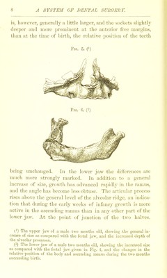
 No text description is available for this image
 No text description is available for this image
 No text description is available for this image