Outlines of the nerves : with short descriptions : designed for the use of medical students / by John Neill.
- John Neill
- Date:
- 1852
Licence: Public Domain Mark
Credit: Outlines of the nerves : with short descriptions : designed for the use of medical students / by John Neill. Source: Wellcome Collection.
Provider: This material has been provided by the National Library of Medicine (U.S.), through the Medical Heritage Library. The original may be consulted at the National Library of Medicine (U.S.)
39/52
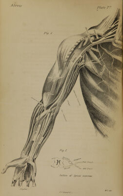
![TIIF. NERVES. SUB-OCCIPITAL X E H V E . — This nerve is exceedingly small, and generally arises by a single root from the spinal marrow. It passes out of the vertebral cavity between the occiput and the atlas, and supplies the muscles on the back of the neck and head. [Plate VI.; and Plate IV., Fig. 1.] THREE SUPERIOR CERVICAL NERVES.—After escaping through the intervertebral foramina, they divide into anterior and pos- terior trunks. The posterior trunks are spent upon the muscles upon the back of the spinal column. The anterior trunks form a Plexus, branches from which supply the muscles of the neck ; and cutaneous branches are distributed to the integuments. The Great Auricular branch passes behind the ear. PHRENIC NERVE. — This nerve is formed from the second and third cervical, descends upon the front of the scalenus amicus muscle, and then enters the thorax through the superior medi- astinum ; passing over the pericardium is distributed to the dia- phragm. FOUR INFERIOR CERVICAL NERVES. Plate VII. —The pos- terior branches are distributed to the muscles and back. The anterior branches are larger, and descend between the scalenus anticus and scalenus medius muscles, above the subclavian artery, to form the brachial plexus. BRACHIAL OR AXILLARY PLEXUS. —This plexus is formed by the junction of the four inferior cervical nerves and the first dorsal, and extends from the scaleni muscles to the neck of the humerus, surrounding the axillary artery. It gives off the fol- lowing nerves, viz: Scapular. — It passes backwards over the shoulder, through the coro- coid notch of the scapula, and is distributed to the spinati mus- cles. Subscapular. — These are usually three in number. Thej are distri- buted to the subscapularis and teres muscles.](https://iiif.wellcomecollection.org/image/b21143584_0039.jp2/full/800%2C/0/default.jpg)