Volume 1
A textbook of human physiology / / translated from [the] 7th German edition by William Stirling.
- Landois, Leonard
- Date:
- 1891
Licence: Public Domain Mark
Credit: A textbook of human physiology / / translated from [the] 7th German edition by William Stirling. Source: Wellcome Collection.
Provider: This material has been provided by Royal College of Physicians, London. The original may be consulted at Royal College of Physicians, London.
48/602 (page 8)
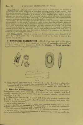
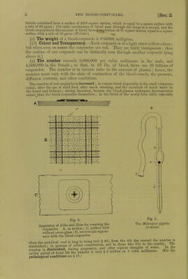
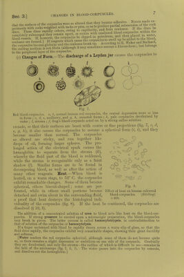
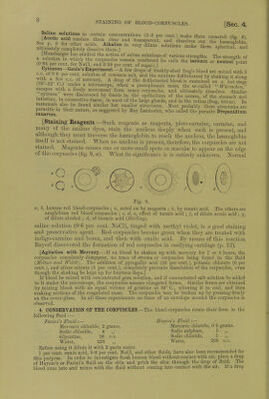
![fAcentio8Si011S,in <ei,tam 7nceilt™ti0»s (2-3 per cent.) make them erenated (fig. 6). See p S S „fW erSn-^llemA1vev1' tlan'si;a,,e,,t' alld di*solves out the l^noglobin uitin!ijyirteiivo^om very te solutions nmke them spherica!-aud [Hamburger has studied the action of saline solutions of various strengths. The strength of foS iS011 * 1(S #e Wfi remain unaltered 116 calls the isotonic 01 itoS oiu (0 64 per cent, for NaCl, and 5'59 per cent, of su«ar) ] 1 Cytozoon-Gaule's Experiment.-A few dropsof freshly-shed frog's blood are mixed with 5 c.c. ol 0-6 per cent solution of common salt, and the mixture defibrinated by shaking it alone sr? so° n\ C-C- i°f merc.ui7- A drop of the defibrinated blood is examined on a hot stage (6V -61 u under a microscope, when a protoplasmic mass, tbe so-called Wiirmchcn escapes with a lively movement from many corpuscles, and ultimately dissolves. Similar cytozoa were discovered by Gaule in the epithelium of the cornea, of the stomach and intestine, in connective-tissue, in most of the large glands, and in the retina (frog, triton) In mammals also he found similar but smaller structures. Most probably these structures are parasitic in their nature, as suggested by Ray Lankester, who called the parasite Drepanidium ranarum. [Staining Reagents.—Such reagents as magenta, picro-carmine, carmine, and many of the aniline dyes, stain the nucleus deeply when such is present, and although they must traverse the haemoglobin to reach the nucleus, the haemoglobin itself is not stained. When no nucleus is present, therefore, the corpuscles are not stained. Magenta causes one or more small spots or maculae to appear on the edge of the corpuscles (fig. 9, a). What its significance is is entirely unknown. Normal Fig. 9. «, b, human red blood-corpuscles ; a, acted on by magenta ; 6, by tannic acid. The others are amphibian red blood-corpuscles ; c, cl, e, effect of tannic acid ; /, of dilute acetic acid ; g, of dilute alcohol ; cl, of boracic acid [Stirling). saline solution (06 per cent. NaCl), tinged with methyl violet, is a good staining and preservative agent. Eed corpuscles become green when they are treated with indigo-carmine and borax, and then with oxalic acid. By means of this reaction Bayerl discovered the formation of red corpuscles in ossifying cartilage (p. 12). [Agitation with Mercury.—If ox blood be shaken up with mercury for 7 or 8 hours, the corpuscles completely disappear, no trace of stroma or corpuscles being found in the fluid (Meltzcr and Welch). The addition of pyrogallic acid (20 percent.), potassic chlorate (6 per cent.), and silver nitrate (3 per cent.), completely prevents dissolution of the corpuscles, even though the shaking be kept up for fourteen days.] If blood be mixed with concentrated gum solution, and if concentrated salt solution be added to it under the microscope, the corpuscles assume elongated forms. Similar forms are obtained by mixing blood with an equal volume of gelatine at 36° C, allowing it to cool, and then making sections of the coagulated mass. The corpuscles may be broken up by pressing firmly on the cover-glass. In all these experiments no trace of an envelope around the corpuscles is observed. 4. CONSERVATION OF THE CORPUSCLES. —The blood-corpuscles retain their form in the following fluid :— Pacini's Fluid :— Hayem's Fluid ;— Mercuric chloride, 2 grams. Mercuric chloride, 0'5 grams. Sodic chloride, 4 ,, Sodic sulphate, 5 „ Glycerine, 26 c.c. Sodic chloride, 1 ,, Water, 226 ,, Water, 200 c.c. Before using it dilute it with 2 parts water. 1 per cent, osmic acid, 06 per cent. NaCl, and other fluids, have also been recommended for this purpose. In order to investigate fresh human blood without contact with air, place a drop of Hayem's or Pacini's fluid on the skin and prick the skin through the drop of fluid. The blood runs into aud mixes with the fluid without coming into contact with the air. If a drop](https://iiif.wellcomecollection.org/image/b24757342_0001_0048.jp2/full/800%2C/0/default.jpg)