Volume 1
A textbook of human physiology / / translated from [the] 7th German edition by William Stirling.
- Landois, Leonard
- Date:
- 1891
Licence: Public Domain Mark
Credit: A textbook of human physiology / / translated from [the] 7th German edition by William Stirling. Source: Wellcome Collection.
Provider: This material has been provided by Royal College of Physicians, London. The original may be consulted at Royal College of Physicians, London.
54/602 (page 14)
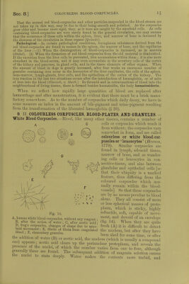
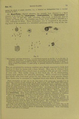
![[According to Bizzozero, there is no evidence to show that the white corpuscles are precursors of the red; the red corpuscles are derived from special corpuscles (erythroblasts), and so are the white (leucoblasts). The red ones seem to he formed within the blood-vessels of the red marrow and the colourless ones in the extra-vascular parts of the marrow. The red corpuscles are formed by the mitotic division of pre-existing cells, which are quite different from the Fie. 13. colourless corpuscles; their protoplasm A, Red blood-corpuscle of a chick undergoing is never granular, but almost always mitotic division at 5th-6th day of incubation homogeneous, never colourless, but B, red blood-corpuscle of frog dividing; Y slightly tinged by haemoglobin ; they shows a thin colourless thread of protoplasm never exhibit the lively amoel)ni(l move. still connecting the two daughter corpuscles. , £ ,1 , J , Tf 6 y ments of the white corpuscles. If the red marrow of any of the classes of the vertebrata be examined, especially after repeated haemorrhages—there will always be found numerous erythroblasts under- going mitosis (fig. 13).] [In all classes of the Vertebrata, then, the red marrow is the great seat of the formation of red corpuscles during adult life. But how is it during the develop- ment of the young animals 1 It is not necessary to assume that the red are derived from the colourless corpuscles. If we study the fate of the red corpuscles we find that their presence is not due absolutely to any one organ. In the first phases of embryonic life, the red corpuscles develop and divide within the whole vascular system. At a later period this ceases and they are developed in the liver and spleen ; at a later period still—in extra-uterine life—and when the bone marrow is greatly developed the blood-forming activity of the liver and spleen is gradually diminished and ceases. But the loss is not absolute in the case of the last organ, as it can again be caused to produce red corpuscles after copious haemorrhage. The blood-plates are in no way concerned in the formation of red corpuscles, they have to do with the coagulation and other vital phenomena of the blood.] [The balance of evidence points to the formation of red blood-corpuscles in extra-uterine life—both in animals with nucleated and in those with non-nucleated corpuscles—by the same process as in embryonic life (i.e., by indirect division or mitosis of a typical cellular element, which during extra-uterine life is chiefly found in the marrow of bone (Bizzozero).] 8. DECAY OF THE RED BLOOD-CORPUSCLES. —The blood-corpuscles undergo decay within a limited time, and the liver is regarded as one of the chief organs in which their disintegration occurs, because bile-pigments are formed from haemoglobin, and the blood of the hepatic vein contains fewer red corpuscles than the portal vein. The splenic pulp contains cells which indicate that coloured corpuscles are broken up within it. These are the so-called blood-corpuscle containing cells (§ 103). Quincke's observations go to show that the red corpuscles—which may live from three to four weeks—when about to disintegrate, are taken up by the white blood-corpuscles in the hepatic capillaries, by the cells of the spleen and the bone-marrow, and are stored up chiefly in the capillaries of the liver, in the spleen, and in the marrow of hone. They are transformed partly into coloured, and partly into colourless proteids which contain iron, and are either deposited in a granular form, or arc dissolved. Part of the products of decomposition is used for the formation of new blood-corpuscles in the marrow and in the spleen, and also perhaps in the liver, while a portion of the iron is excreted by the liver in the bile.](https://iiif.wellcomecollection.org/image/b24757342_0001_0054.jp2/full/800%2C/0/default.jpg)