Volume 1
A textbook of human physiology / / translated from [the] 7th German edition by William Stirling.
- Landois, Leonard
- Date:
- 1891
Licence: Public Domain Mark
Credit: A textbook of human physiology / / translated from [the] 7th German edition by William Stirling. Source: Wellcome Collection.
Provider: This material has been provided by Royal College of Physicians, London. The original may be consulted at Royal College of Physicians, London.
573/602 (page 533)
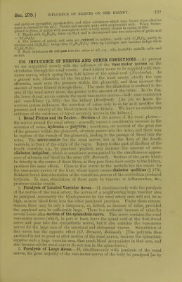
![ONCOMETER. we can measure the variations in the size of a limb, while by the oncograph (07*09, volume) similar variations in the volume of the spleen are measured Wd). Kov and Cohnheim have measured the variations in the volume of the kidney by means of an instrument which consists of two parts, one termed the oncometer or renal plethysmometer, in which the organ is enclosed, while the other part is the registering portion or oncograph. The kidney is enclosed in a kidney-shaped metallic capsule (figs. 347, 348), composed of two halves which move on the hinge, h, to introduce the organ. The renal vessels pass out at a, v. Ine kidney is surrounded with a thin membrane, and between this membrane and the inner surface of the capsule is a space filled with warm oil through the tube, I, which is closed by means of a stop-cock after the space is filled with oil. The kibe T can be in ale to communicate with another tube, Tv leading into a metallic chamber, C, of the oncograph (fig. 349), which is provided with a movable piston, p, attached by a thread to the writing-lever, Z. Any increase in the size of the organ expels oil from the chamber, O, into C, and thus the piston is raised, while a diminution in the size of the kidney diminishes the fluid in C', and the lever falls. The actual volume of the living kidney depends upon the state of distention of its structural elements, upon the amount of lymph in its lymph-spaces, but chiefly upon the amount of blood in its blood-vessels, and this again must depend upon the condition of the non-striped muscles in the renal arteries. When the vessels dilate, the kidney increases in size, and when they contract it contracts, so that we can register on the same revolving cylinder the variations of the volume at the same time that we record the general arterial blood-pressure.] [in the normal circulation through the kidney, the kidney-curve, i.e., the curve of the volume of the kidney, runs parallel with the blood-pressure curve, and show s B.I '/i MIN Fig. 350. B. P., Blood-pressure curve ; K., curve of the volume of the kidney ; T, time curve : intervals indicate a quarter of a minute ; A, abscissa {Stirling, after Roy). the large respiratory undulations, as well as the smaller elevations due to the systole of the heart (fig. 350). In this respect it differs sharply from a spleen-curve (fig. 140). Usually, when the blood-pressure falls, the kidney-curve sinks, and when the blood-pressure rises the volume of the kidney increases. When the blood-pressure curve is complicated by Traube-Hering waves (§ 85) the opposite effect is produced on the kidney-curve ; the highest blood-pressure corresponds to the smallest size of the kidney, and conversely. This is due to the fact that, when these curves occur, all the small arterioles, including those in the kidney, are contracted. A kidney placed in an oncometer secretes urine like a kidney under natural conditions.] [Arrest of the respiration in a curarised animal produces a rapid and great diminution of the, volume of the kidney, caused by the venous blood stimulating the vaso-motor centres, and thus contracting the small arterioles, including those of the kidney. This result occurs whether one or both splanehnics arc divided, proving that all the vaso-motor nerves of the kidney do not reach it through the splaneh- nics. When all the renal nerves at the hilum are divided, arrest of the respiration](https://iiif.wellcomecollection.org/image/b24757342_0001_0573.jp2/full/800%2C/0/default.jpg)