Volume 1
A textbook of human physiology / / translated from [the] 7th German edition by William Stirling.
- Landois, Leonard
- Date:
- 1891
Licence: Public Domain Mark
Credit: A textbook of human physiology / / translated from [the] 7th German edition by William Stirling. Source: Wellcome Collection.
Provider: This material has been provided by Royal College of Physicians, London. The original may be consulted at Royal College of Physicians, London.
576/602 (page 536)
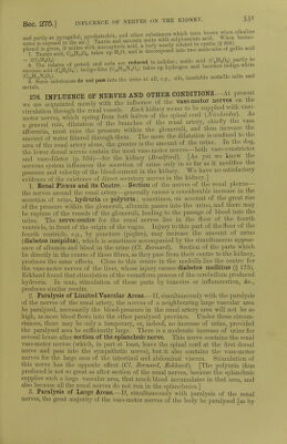
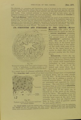
![The alkaloids, i.e., ptomaines and leucomaiucs, seem to be formed by the action of vegetable organisms in the intestine, whence they are absorbed into the blood, and pass into the urine (§ 116). This, however, is denied by some observers, who state that normal urine is free from such bodies. Urine rendered colourless by charcoal loses half its toxic power, and the poisonous substance is not volatile, and even resists boiling. These alkaloids arc increased in the urine in typhoid lever, pneumonia, but not in diabetes.] Uric Acid Diathesis.—When too much nitrogenous food, too much of any alcoholic fluid is persistently used, and little muscular exercise taken, especially if the respiratory organs are in- terfered with, uric acid may not unfrequently accumulate, in the blood (Garrod). It may be deposited in the joints and their ligaments, especially in the foot and hand, giving rise to pain- ful inflammation, and forming gout-stones or chalk-stones [which are acid-urates]. The heart, liver, and kidneys are rarely affected. The tissues near these deposits undergo necrosis. 278. STEUCTTJEE AND FUNCTIONS Adventltin. m ' v • o o.. ■■ fljlV OF THE UEE TEE.—Mucous Membrane.—The pelvis of the kidney and the ureter are lined by a mucous membrane, consisting of connective-tissue, and covered with several layers of stratified transitional epithelium (fig. 352). The cells are of various shapes, those of the lowest layer being usually more or less spheri- cal and small, while many of the cells in the upper layers are ir- regular in shape, often with long processes passing into the deeper layers. Sub-mucosa.—Under the epi- thelium there is a layer of adenoid tissue (Hamburger, Chiari), which may contain small lymph-follicles [embedded in loose connective- tissue]. In the pelvis of the kidney and ureter there are a few small mucous glands lined Fig. 351. Transverse section of the lower part of human ureter, x 15. e, epithelium ; t, tunica propria ; s, sub- mucosa ; I and r, longitudinal and circular fibres. by a single layer of columnar epithelium (Unruh, Egli). _ The muscular coat consists of an inner somewhat stronger layer of longitudinal Cylindrical non-striped fibres, and an outer circular oells' layer (fig. 351). In the lowest third of the ureter there are in addition a number of scattered muscular fibres. All these layers are surrounded and supported by connec- tive-tissue. The outer layers of the connec- tive-tissue form an outer coat or adventitia, which contains the large vessels and nerves. The various coats of the ureter can be followed up to the pelvis of the kidney, and to its calices. The papilla- are covered only by the mucous membrane, while the muscular layer ceases at the apex of the pyramids, where they are disposed circu- larly, to form a kind of sphincter muscle for each papilla (Henle). Lcucocvte. Tunica propria. Vertical section of the mucous membrane of a human bladder. 3ESKIS5S»2 =2* The nerves are fibres, with numerous &.](https://iiif.wellcomecollection.org/image/b24757342_0001_0576.jp2/full/800%2C/0/default.jpg)