Volume 1
A textbook of human physiology / / translated from [the] 7th German edition by William Stirling.
- Landois, Leonard
- Date:
- 1891
Licence: Public Domain Mark
Credit: A textbook of human physiology / / translated from [the] 7th German edition by William Stirling. Source: Wellcome Collection.
Provider: This material has been provided by Royal College of Physicians, London. The original may be consulted at Royal College of Physicians, London.
63/602 (page 23)
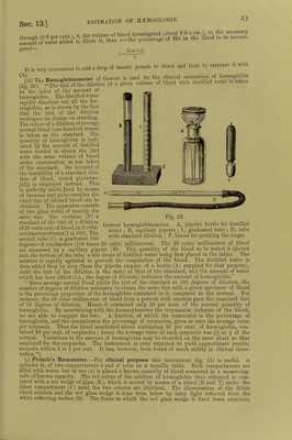
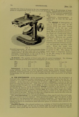
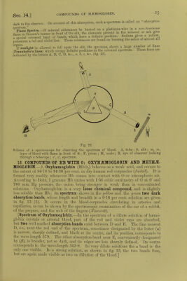
![ESTIMATION OF HAEMOGLOBIN. gated— k{w+b) It is very convenient to add a drop of caustic potash to blood and then to saturate it with l',^ m. w^ncrlnhmometer of Gowers is used for the clinical estimation of haemoglobin m) ^^^ZtSllo, oi a given volume of blood with distilled water is taken CO. (fig. 20). as the index of the amount ot haemoglobin. The distilled water rapidly dissolves out all the hae- moglobin, as is shown by the fact that the tint of the dilution undergoes no change on standing. The colour of a dilution of average normal blood (one hundred times) is taken as the standard. The quantity of haemoglobin is indi- cated by the amount of distilled water needed to obtain the tint with the same volume of blood under examination as was taken of the standard. On account of the instability of a standard dilu- tion of blood, tinted glycerine- jelly is employed instead. This is perfectly stable,fand by means of carmine and picro-carmine the exact tint of diluted blood can be obtained. The apparatus consists of two glass tubes of exactly the same size. One contains (D) a standard of the tint of a dilution of 20 cubic mm. of blood, in 2 cubic centimetresofwater(l in 100). The second tube (0) is graduated 100 Fig. 20. Gowers' haemoglobinonieter. A, pipette bottle for distilled water ; B, capillary pipette ; C, graduated tube ; D, tube with standard dilution ; F, lancet for pricking the finger. degrees = 2 centimetres (100 times 20 cubic millimetres). The 20 cubic millimetres of blood are measured by a capillary pipette (B). This quantity of the blood to be tested is ejected into the bottom of the tube, a few drops of distilled water being first placed in the latter. The mixture is rapidly agitated to prevent the coagulation of the blood. The distilled water is then added drop by drop (from the pipette stopper of a bottle (A) supplied for that purpose), until the tint of the dilution is the same as that of the standard, and the amount of water which has been added {i.e., the degree of dilution) indicates the amount of haemoglobin. Since average normal blood yields the tint of the standard at 100 degrees of dilution, the number of degrees of dilution necessary to obtain the same tint with a given specimen of blood is the percentage proportion of the haemoglobin contained in it, compared to the normal. For instance, the 20 cubic millimetres of blood from a patient with anaemia gave the standard tint of 30 degrees of dilution. Hence it contained only 30 per cent, of the normal quantity of haemoglobin. By ascertaining with the haemacytometer the corpuscular richness of the blood, we are able to compare the two. A fraction, of which the numerator is the percentage of haemoglobin, and the denominator the percentage of corpuscles, gives at once the average value per corpuscle. Thus the blood mentioned above containing 30 per cent, of haemoglobin, con- tained 60 per cent, of corpuscles ; hence the average value of each corpuscle was or £ of the normal. Variations in the amount of haemoglobin may be recorded on the same chart as that employed for the corpuscles. The instrument is only expected to yield approximate results, accurate within 2 or 3 per cent. It has, however, been found of much utility in clinical obser- vation.] (e) Fleischl's Hsomometer.—For clinical purposes this instrument (fig. 21) is useful. A cylinder G, of two compartments a and a' rests on a metallic table. Both compartments are filled with water, but in one {a) is placed a known quantity of blood measured in a measuring- tube of known capacity. The red colour of the solution of haemoglobin thus obtained is com- pared with a red wedge of glass (K), which is moved by means of a wheel (It and T) under the other compartment («') until the two colours are identical. The illumination of the dilute blood solution and the red glass wedge is done from below by lamp light reflected from the white reflecting surface (S). The frame in which the red glass wedge is fixed bears numbers,](https://iiif.wellcomecollection.org/image/b24757342_0001_0063.jp2/full/800%2C/0/default.jpg)