Möller's operative veterinary surgery / translated and edited from the second enlarged and improved edition of 1894 by Jno. A.W. Dollar.
- Date:
- 1895
Licence: Public Domain Mark
Credit: Möller's operative veterinary surgery / translated and edited from the second enlarged and improved edition of 1894 by Jno. A.W. Dollar. Source: Wellcome Collection.
Provider: This material has been provided by the Royal College of Physicians of Edinburgh. The original may be consulted at the Royal College of Physicians of Edinburgh.
674/768 page 646
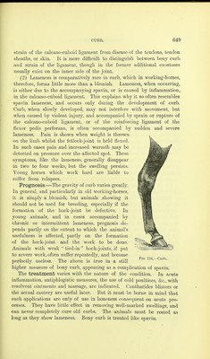
![many have remained for years free from lameness. Failing success by this method, the pointed cautery may sometimes he found of more service. Its use is without danger, the large exostoses preventing it entering the joint. Klemm's method of dividing the flexor metatarsi muscle 3 or 4 inches above the hock-joint is, in my experience, useless. If divided completely, lameness follows similar to that after rupture of the muscle (p. 619). By giving four to six weeks' rest, the joint may become auchylosed, and lameness disappear, but this often fails to occur. Par- tial section sometimes disguises the stringhalt-like lameness, but cannot cure the disease of the joint, and the owner generally returns after an interval to submit the horse to further treatment. In the Prussian army the actual cautery has, during the last few years, been largely used in treating spavin, the successes numbering about 60 per cent. The method recommended consists in perforating the bursa with a pyriform iron, which is passed into the bone. Peri- osteotomy proved of less value. It must, however, be remembered that in no other disease are diagnostic errors so frequent as here, for even the most careful examination often leads to no definite conclusion. Serious methods of treatment are only applied to serious conditions, i.e., to cases in which disease has made extensive progress, whilst the milder cases are blistered or fired; especially in the army, where firing and blistering are greatly relied on. As Hering pointed out, neurotomy is not of much use in spavin. Section of the tibial nerve above the hock, or of the cutaneous nerves, does not remove the pain or lameness. This is all the more unfortunate, as all the above-described methods of treatment are at times unsuccess- ful, and leave the horse permanently lame. The shoeing is of some importance. Klemm recommends raising the heels, and giving long quarters and a short toe, a suggestion I can fully support. Koster also recommended shortening the toe of the foot before treatment, and using long, wide shoes, with heels and toe-pieces. VI-ENLARGEMENTS ON THE OUTER SURFACE OF THE HOCK/ Ger. Eehbein. The above title includes all circumscribed thickenings on the outside of the hock-joint. They may be situated in the ligamentous apparatus, 1 A literal translation of the German title would have no meaning to English ears, and yet no specific title exists in English. I can best explain Kehbein by describing it as spavin on the outer surface of the hock.—[Tkansl.]](https://iiif.wellcomecollection.org/image/b2193986x_0674.jp2/full/800%2C/0/default.jpg)